

Topographical Central Island-Like Pattern After 24 Hrs of Continuous Intraocular Pressure Monitoring with a Contact Lens Sensor
- PMID: 32104102
- PMCID: PMC7023676
- DOI: 10.2147/IMCRJ.S232659
Topographical Central Island-Like Pattern After 24 Hrs of Continuous Intraocular Pressure Monitoring with a Contact Lens Sensor
Abstract
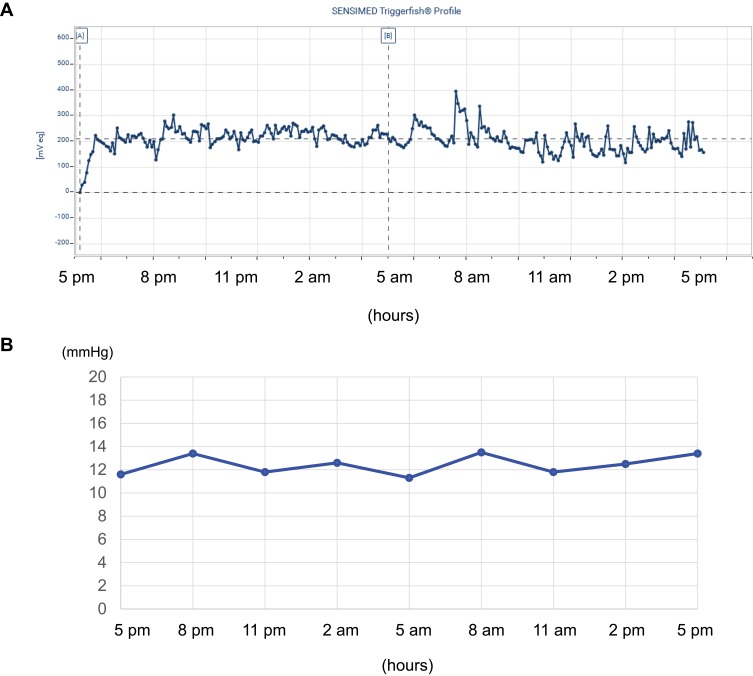
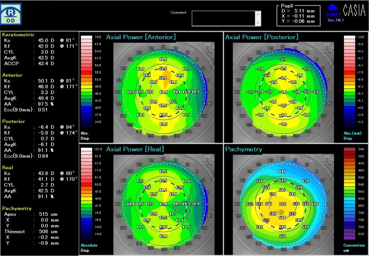
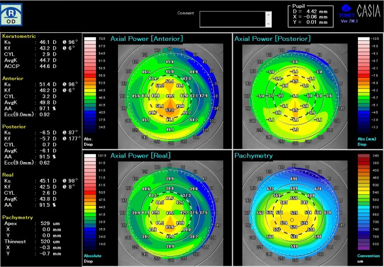
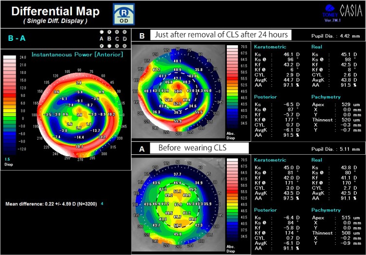
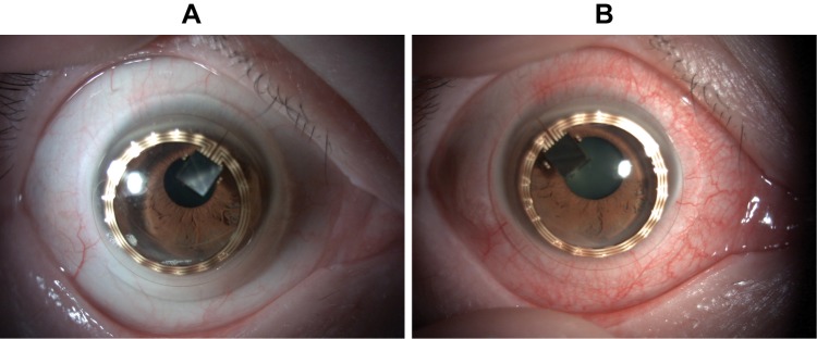
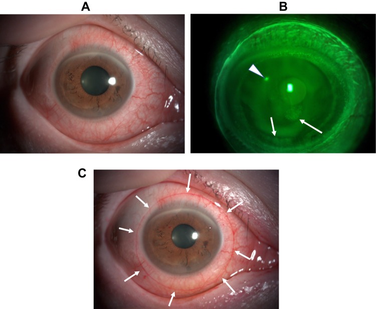
With development of the contact lens sensor (CLS), it has become possible to monitor the intraocular pressure (IOP) for 24 hrs continuously. Wearing of CLS often brings blurred vision with transient aggravation of myopia and changes in corneal shape. The author, a 51-year-old man with myopic astigmatism, wore a CLS for 24 hrs on the right eye, and the fellow eye served as a contra-lateral control eye. After wearing, his corrected visual acuity on the right eye decreased from 20/16 to 20/25 with blurred vision, and subjective spherical power and cylindrical power aggravated. Topographical analysis revealed that the instantaneous power increased on the central cornea but decreased on the mid-peripheral cornea. Differential instantaneous map of pre- and post-wearing CLS showed a specific pattern similar to the central island pattern, which is known as the results of steeper fitting of the orthokeratology lens. A surface imprint was observed on the bulbar conjunctiva, corresponding to the edge of the contact lens. These findings seemed due to orthokeratological effects by the steeper fitting of CLS. All of them resolved within 24 hrs after the removal of the CLS.
Keywords: CLS; central island; contact lens sensor; orthokeratological effects; topography; triggerfish.
© 2020 Toshida.
Conflict of interest statement
Dr Hiroshi Toshida reports grants from the Ministry of Education, Culture, Sports, Science and Technology, during the conduct of the study and outside the submitted work. The author reports no other conflicts of interest in this work.
Figures
Similar articles
-
Transient changes in refractive error and corneal tomography after 24-h continuous monitoring of intraocular pressure patterns with a contact lens sensor.Jpn J Ophthalmol. 2020 Mar;64(2):127-133. doi: 10.1007/s10384-020-00723-6. Epub 2020 Feb 13. Jpn J Ophthalmol. 2020. PMID: 32056036
-
[OPTIC PROPERTIES OF MYOPIC CORRECTION BY ORTHOKERATOLOGY CONTACT LENSES (A CASE REPORT)].Cesk Slov Oftalmol. 2017 Spring;73(1):17-23. Cesk Slov Oftalmol. 2017. PMID: 28639449 Czech.
-
Corneal thickness after overnight wear of an intraocular pressure fluctuation contact lens sensor.Acta Ophthalmol. 2012 Nov;90(7):e534-9. doi: 10.1111/j.1755-3768.2012.02495.x. Epub 2012 Sep 13. Acta Ophthalmol. 2012. PMID: 22974389 Clinical Trial.
-
[Quantitative assessment of quality of vision].Nippon Ganka Gakkai Zasshi. 2004 Dec;108(12):770-807; discussion 808. Nippon Ganka Gakkai Zasshi. 2004. PMID: 15656087 Review. Japanese.
-
Outcome, influence factor and development of CLS measurement in continuous IOP monitoring: A narrative review.Cont Lens Anterior Eye. 2021 Aug;44(4):101376. doi: 10.1016/j.clae.2020.10.006. Epub 2020 Oct 19. Cont Lens Anterior Eye. 2021. PMID: 33092960 Review.
Cited by
-
Advancements in Wearable and Implantable Intraocular Pressure Biosensors for Ophthalmology: A Comprehensive Review.Micromachines (Basel). 2023 Oct 9;14(10):1915. doi: 10.3390/mi14101915. Micromachines (Basel). 2023. PMID: 37893352 Free PMC article. Review.
References
-
- Liu JH, Kripke DF, Hoffman RE, et al. Nocturnal elevation of intraocular pressure in young adults. Invest Ophthalmol Vis Sci. 1998;39(13):2707–2712. - PubMed
Publication types
LinkOut - more resources
Full Text Sources
