. 2020 Mar;19(3):1903-1907.
doi: 10.3892/etm.2020.8423. Epub 2020 Jan 3.

Abscopal effect following nivolumab induction in a patient with metastatic renal cell carcinoma-unique pathological features of the primary specimen: A case report

Affiliations

Abscopal effect following nivolumab induction in a patient with metastatic renal cell carcinoma-unique pathological features of the primary specimen: A case report

Kanta Hori et al. Exp Ther Med. 2020 Mar.

Abstract

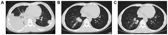
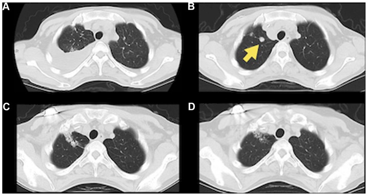
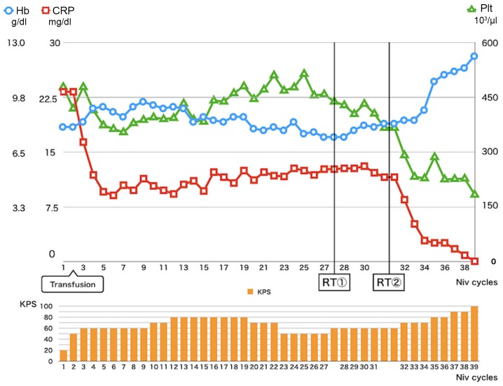
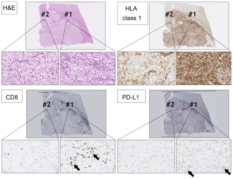
The case of a patient with metastatic renal cell carcinoma who exhibited the abscopal effect following treatment by anti-programmed death-1 (PD-1) antibody is presented. A 40-year-old woman was diagnosed with an 8.2-cm renal tumor without distant metastases, and radical nephrectomy was subsequently performed. Pathological examination revealed a clear cell renal cell carcinoma. At 3 months after surgery, the patient developed one lung metastasis. Following treatment with interferon and three types of tyrosine kinase inhibitors, anti-PD1 antibody (nivolumab) was started. During the treatment, para-aortic/supraclavicular lymph nodes and several lung lesions remained, although other lesions decreased markedly. The patient was subsequently treated by palliative radiotherapy to the para-aortic and supraclavicular lymph nodes for pain control. After the radiotherapy, the lung lesions previously refractory to nivolumab started to decrease, probably due to an abscopal effect. Additionally, the laboratory data and Karnofsky Performance Status improved. Histological re-examination of the primary lesion revealed heterogeneity of the immunological microenvironment, which may be associated with the heterogeneity of treatment sensitivity.

Keywords: abscopal effect; anti-PD1 antibody; cytotoxic T lymphocyte; cytotoxic T lymphocytes; immune checkpoint inhibitor; radiation therapy; renal cell carcinoma.


Figures

Figure 1.
Figure 1.
Images of the lung nodules after nivolumab induction. (A) Pretreatment computed tomography reveals increases in the number and size of nodules in the lung lobes with pleural effusion. After (B) 6 and (C) 26 cycles, most lung nodules had shrunk, and the pleural effusion had completely resolved.
Figure 2.
Figure 2.
Images of lymph nodes after nivolmab induction and radiotherapy. (A and B) Right supraclavicular lymph node (yellow arrow) was enlarged after 26 cycles of nivolumab. (C) After 4 months following radiotherapy, an objective response was observed in the right supraclavicular lymph node(yellow arrow). (D and E) Para-aortic lymph node (yellow arrow) was enlarged after 26 cycles of nivolumab. (F) After 8 months following radiotherapy, an objective response was observed in the para-aortic lymph node (yellow arrow).
Figure 3.
Figure 3.
Images of abscopal effect on the nivolumab-resistant lung nodules. (A and B) One nodule (yellow arrow) in the right lung had grown after nivolumab induction. After (C) 1 month (C and D) 6 months following radiotherapy to the lymph nodes, a treatment response was seen in the lung lobe.
Figure 4.
Figure 4.
Transition of laboratory data and KPS during the Following the Niv induction, CRP improved partially. At 3 months after the RT, CRP, Hb and Plt values were normalized. KPS also improved from 50 to 100. Niv, nivolumab; CRP, C-reactive protein; RT, radiotherapy; Hb, hemoglobin; Plt, platelet; KPS, Karnofsky Performance Status.
Figure 5.
Figure 5.
Histological analysis of the primary RCC lesion. RCC sections were stained with H&E, anti-CD8 antibody (arrows), anti-HLA class 1 antibody, and anti-PD-L1 antibody (arrows). Magnification, ×2 or ×400. There were two staining patterns in RCC lesions #1 and #2. RCC, renal cell carcinoma; H&E, hematoxylin and eosin; HLA, human leukocyte antigen; PD-L1, programmed cell death ligand 1.

References

    1. Mole RH. Whole body irradiation; radiobiology or medicine? Br J Radiol. 1953;26:234–241. doi: 10.1259/0007-1285-26-305-234. - DOI - PubMed
    1. Formenti SC, Demaria S. Combining radiotherapy and cancer immunotherapy: A paradigm shift. J Natl Cancer Inst. 2013;105:256–265. doi: 10.1093/jnci/djs629. - DOI - PMC - PubMed
    1. Galluzzi L, Chan TA, Kroemer G, Wolchok JD, López-Soto A. The hallmarks of successful anticancer immunotherapy. Sci Transl Med. 2018;10:eaat7807. doi: 10.1126/scitranslmed.aat7807. - DOI - PubMed
    1. Sun M, Shariat SF, Cheng C, Ficarra V, Murai M, Oudard S, Pantuck AJ, Zigeuner R, Karakiewicz PI. Prognostic factors and predictive models in renal cell carcinoma: A contemporary review. Eur Urol. 2011;60:644–661. doi: 10.1016/j.eururo.2011.06.041. - DOI - PubMed
    1. Shinohara N, Abe T. Prognostic factors and risk classifications for patients with metastatic renal cell carcinoma. Int J Urol. 2015;22:888–897. doi: 10.1111/iju.12858. - DOI - PubMed