. 2020 Mar;19(3):2282-2290.
doi: 10.3892/etm.2019.8403. Epub 2019 Dec 31.

Cystatin C promotes cognitive dysfunction in rats with cerebral microbleeds by inhibiting the ERK/synapsin Ia/Ib pathway

Affiliations

Cystatin C promotes cognitive dysfunction in rats with cerebral microbleeds by inhibiting the ERK/synapsin Ia/Ib pathway

Guangna Yu et al. Exp Ther Med. 2020 Mar.

Abstract

Although higher serum level of cystatin C (CysC) was observed in patients with cerebral microbleeds, its associated role in the disease has not been elucidated. In this work, a rat model of cerebral microbleeds was created with the aim of investigating effects of CysC on cognitive function in rats with cerebral microbleeds and the underlying mechanism. Serum samples of patients with cerebral microbleeds and healthy people of the same age were collected. Levels of cystatin C expression in these samples were measured using CysC kits. Moreover, 48 spontaneously hypertensive rats (SHRs) bred under specific pathogen-free (SPF) conditions were randomly divided into 4 groups: sham surgery control group (sham), model group (CMB), model + empty vector control group (CMB + vehicle), and model + cystatin C overexpression group (CMB + CysC). Expression levels of CysC in hippocampus of rats in each group were measured by western blot analysis. The Y-maze was used to evaluate cognitive function of rats. Hippocampal long-term potentiation (LTP) in rats was assessed by the electrophysiological assay. Alterations in levels of p-ERK1/2 and p-synapsin Ia/b proteins associated with cognitive function were identified by western blot analysis. The serum levels of CysC in patients with cerebral microbleeds were significantly upregulated (P<0.001). After injection of CysC, its expression levels in rat hippocampus were significantly increased (P<0.001), which enhanced the decline in learning and memory function, as well as the decrease of LTP in the rat model of cerebral microbleeds (P<0.001). Western blot results showed that injection of CysC further reduced the levels of p-ERK1/2 and p-synapsin Ia/b in the rat model of microbleeds (P<0.001). CysC was up regulated in serum of patients with cerebral microbleeds. It promoted cognitive dysfunction in rats with microbleeds by inhibiting ERK/synapsin Ia/Ib pathway.

Keywords: brain microbleeds; cognitive function; cystatin C.


Figures

Figure 1.
Figure 1.
CysC expression levels in serum of healthy subjects and patients with cerebral microbleeds. Compared with healthy controls, the expression level of CysC in serum of patients with cerebral microbleeds increased significantly. ***P<0.001. CysC, cystatin C.
Figure 2.
Figure 2.
Expression of CysC in hippocampus of rats in each group: (A) Western blot image of CysC expression in rat hippocampus. (B) Bar charts indicating CysC expression in hippocampus of rats in each group. *P<0.05, ***P<0.001 vs. the sham group; ###P<0.001 vs. the CMB group; n=6 per group. CysC, cystatin C; CMB, cerebral microbleeds.
Figure 3.
Figure 3.
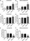
Y-maze test: (A) Number of incorrect reactions in the learning test. (B) Number of incorrect reactions in the memory test. (C) Total reaction time in the learning test. (D) Total reaction time in the memory test. (E) Active avoidance rate in the learning test. (F) Active avoidance rate in the memory test. **P<0.01, ***P<0.001 vs. the sham group; ##P<0.01, ###P<0.001 vs. the CMB group; n=6 per group. CMB, cerebral microbleeds.
Figure 4.
Figure 4.
LTP recordings in each group: (A) Field excitatory postsynaptic potential (fEPSP) amplitudes; (B) Average fEPSP amplitudes in the last 15 min. ***P<0.001 vs. the sham group; ###P<0.001 vs. the CMB group; n=6 per group. LTP, long-term potentiation; CMB, cerebral microbleeds.
Figure 5.
Figure 5.
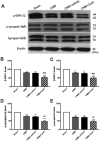
Expression of p-ERK1/2 and p-synapsin in each group: (A) Western blot analysis of p-ERK1/2 and p-synapsinIa/b expression. (B and C) Bar charts indicating p-ERK1/2 expression. (D and E) Bar charts indicating phosphorylation levels of synapsin Ia/b (Ser 549). **P<0.01, ***P<0.001 vs. the sham group; ##P<0.01, ###P<0.001 vs. the CMB group; n=6 per group. CMB, cerebral microbleeds. ERK, extracellular signal-regulated kinase.

Similar articles

Cited by

References

    1. Moulin S, Cordonnier C. Role of cerebral microbleeds for intracerebral haemorrhage and dementia. Curr Neurol Neurosci Rep. 2019;19:51. doi: 10.1007/s11910-019-0969-0. - DOI - PubMed
    1. Cordonnier C, Al-Shahi Salman R, Wardlaw J. Spontaneous brain microbleeds: Systematic review, subgroup analyses and standards for study design and reporting. Brain. 2007;130:1988–2003. doi: 10.1093/brain/awl387. - DOI - PubMed
    1. Daugherty AM, Raz N. Incident risk and progression of cerebral microbleeds in healthy adults: A multi-occasion longitudinal study. Neurobiol Aging. 2017;59:22–29. doi: 10.1016/j.neurobiolaging.2017.07.003. - DOI - PMC - PubMed
    1. Vernooij MW, van der Lugt A, Ikram MA, Wielopolski PA, Niessen WJ, Hofman A, Krestin GP, Breteler MM. Prevalence and risk factors of cerebral microbleeds: The Rotterdam Scan Study. Neurology. 2008;70:1208–1214. doi: 10.1212/01.wnl.0000307750.41970.d9. - DOI - PubMed
    1. Charidimou A, Imaizumi T, Moulin S, Biffi A, Samarasekera N, Yakushiji Y, Peeters A, Vandermeeren Y, Laloux P, Baron JC, et al. Brain hemorrhage recurrence, small vessel disease type, and cerebral microbleeds: A meta-analysis. Neurology. 2017;89:820–829. doi: 10.1212/WNL.0000000000004259. - DOI - PMC - PubMed