

Copper accumulation and the effect of chelation treatment on cerebral amyloid angiopathy compared to parenchymal amyloid plaques
- PMID: 32104807
- PMCID: PMC8826596
- DOI: 10.1039/c9mt00306a
Copper accumulation and the effect of chelation treatment on cerebral amyloid angiopathy compared to parenchymal amyloid plaques
Abstract
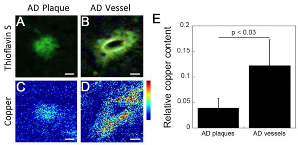
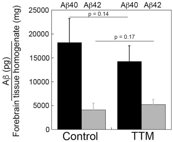
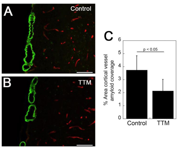
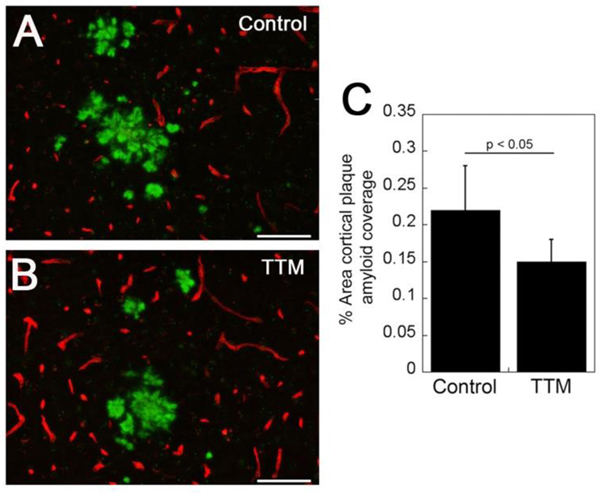
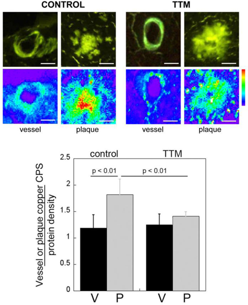
Accumulation of fibrillar amyloid β-protein (Aβ) in parenchymal plaques and in blood vessels of the brain, the latter condition known as cerebral amyloid angiopathy (CAA), are hallmark pathologies of Alzheimer's disease (AD) and related disorders. Cerebral amyloid deposits have been reported to accumulate various metals, most notably copper and zinc. Here we show that, in human AD, copper is preferentially accumulated in amyloid-containing brain blood vessels compared to parenchymal amyloid plaques. In light of this observation, we evaluated the effects of reducing copper levels in Tg2576 mice, a transgenic model of AD amyloid pathologies. The copper chelator, tetrathiomolybdate (TTM), was administered to twelve month old Tg2576 mice for a period of five months. Copper chelation treatment significantly reduced both CAA and parenchymal plaque load in Tg2576 mice. Further, copper chelation reduced parenchymal plaque copper content but had no effect on CAA copper levels in this model. These findings indicate that copper is associated with both CAA deposits and parenchymal amyloid plaques in humans, but less in Tg2576 mice. TTM only reduces copper levels in plaques in Tg2576 mice. Reducing copper levels in the brain may beneficially lower amyloid pathologies associated with AD.
Conflict of interest statement
Conflicts of interest
There are no conflicts to declare.
Figures
Similar articles
-
Evaluation of Copper Chelation Therapy in a Transgenic Rat Model of Cerebral Amyloid Angiopathy.ACS Chem Neurosci. 2023 Feb 1;14(3):378-388. doi: 10.1021/acschemneuro.2c00483. Epub 2023 Jan 18. ACS Chem Neurosci. 2023. PMID: 36651175
-
Human apolipoprotein E4 alters the amyloid-beta 40:42 ratio and promotes the formation of cerebral amyloid angiopathy in an amyloid precursor protein transgenic model.J Neurosci. 2005 Mar 16;25(11):2803-10. doi: 10.1523/JNEUROSCI.5170-04.2005. J Neurosci. 2005. PMID: 15772340 Free PMC article.
-
Early-onset formation of parenchymal plaque amyloid abrogates cerebral microvascular amyloid accumulation in transgenic mice.J Biol Chem. 2014 Jun 20;289(25):17895-908. doi: 10.1074/jbc.M113.536565. Epub 2014 May 14. J Biol Chem. 2014. PMID: 24828504 Free PMC article.
-
Shear-Induced Amyloid Formation in the Brain: I. Potential Vascular and Parenchymal Processes.J Alzheimers Dis. 2016 Sep 6;54(2):457-70. doi: 10.3233/JAD-160027. J Alzheimers Dis. 2016. PMID: 27567812 Free PMC article. Review.
-
Metal chelator decreases Alzheimer beta-amyloid plaques.Neuron. 2001 Jun;30(3):641-2. doi: 10.1016/s0896-6273(01)00330-0. Neuron. 2001. PMID: 11430794 Review.
Cited by
-
Heavy Metal Effects on Biodiversity and Stress Responses of Plants Inhabiting Contaminated Soil in Khulais, Saudi Arabia.Biology (Basel). 2022 Jan 20;11(2):164. doi: 10.3390/biology11020164. Biology (Basel). 2022. PMID: 35205031 Free PMC article.
-
Copper binding and protein aggregation: a journey from the brain to the human lens.RSC Chem Biol. 2023 Oct 17;4(12):974-985. doi: 10.1039/d3cb00145h. eCollection 2023 Nov 29. RSC Chem Biol. 2023. PMID: 38033729 Free PMC article.
-
Heavy Metal-Induced Cerebral Small Vessel Disease: Insights into Molecular Mechanisms and Possible Reversal Strategies.Int J Mol Sci. 2020 May 29;21(11):3862. doi: 10.3390/ijms21113862. Int J Mol Sci. 2020. PMID: 32485831 Free PMC article. Review.
-
A Copper-Binding Peptide with Therapeutic Potential against Alzheimer's Disease: From the Blood-Brain Barrier to Metal Competition.ACS Chem Neurosci. 2025 Jan 15;16(2):241-261. doi: 10.1021/acschemneuro.4c00796. Epub 2024 Dec 26. ACS Chem Neurosci. 2025. PMID: 39723808 Free PMC article.
-
High Redox Status as the Basis for Heavy Metal Tolerance of Sesuvium portulacastrum L. Inhabiting Contaminated Soil in Jeddah, Saudi Arabia.Antioxidants (Basel). 2021 Dec 22;11(1):19. doi: 10.3390/antiox11010019. Antioxidants (Basel). 2021. PMID: 35052523 Free PMC article.
References
-
- Auriel E, Greenberg SM: The pathophysiology and clinical presentation of cerebral amyloid angiopathy. Curr Atheroscler Rep 2012, 14:343–50. - PubMed
-
- Attems J, Jellinger K, Thal DR, Van Nostrand W: Review: sporadic cerebral amyloid angiopathy. Neuropathol Appl Neurobiol 2011, 37:75–93. - PubMed
-
- Rensink AA, de Waal RM, Kremer B, Verbeek MM: Pathogenesis of cerebral amyloid angiopathy. Brain Res Brain Res Rev 2003, 43:207–23. - PubMed
