Review
. 2020 Feb 24:100846.
doi: 10.1016/j.preteyeres.2020.100846. Online ahead of print.

The cell biology of the retinal pigment epithelium

Affiliations
Review

The cell biology of the retinal pigment epithelium

Aparna Lakkaraju et al. Prog Retin Eye Res. .

Abstract

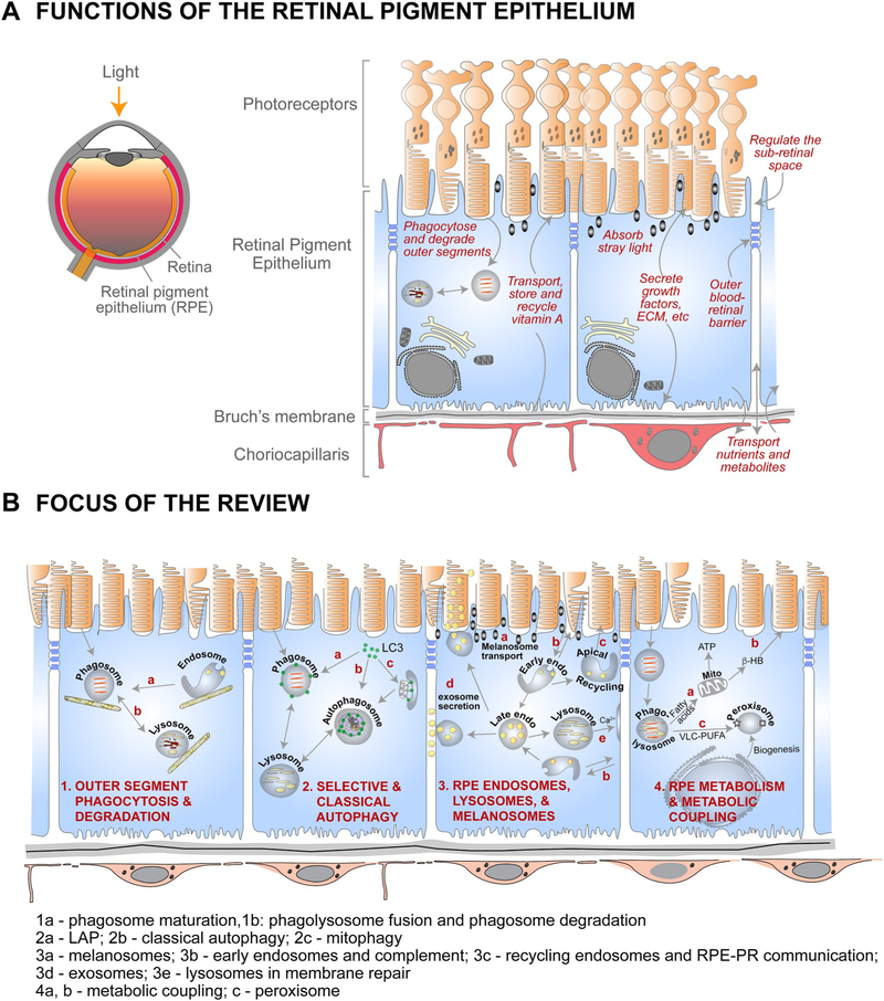
The retinal pigment epithelium (RPE), a monolayer of post-mitotic polarized epithelial cells, strategically situated between the photoreceptors and the choroid, is the primary caretaker of photoreceptor health and function. Dysfunction of the RPE underlies many inherited and acquired diseases that cause permanent blindness. Decades of research have yielded valuable insight into the cell biology of the RPE. In recent years, new technologies such as live-cell imaging have resulted in major advancement in our understanding of areas such as the daily phagocytosis and clearance of photoreceptor outer segment tips, autophagy, endolysosome function, and the metabolic interplay between the RPE and photoreceptors. In this review, we aim to integrate these studies with an emphasis on appropriate models and techniques to investigate RPE cell biology and metabolism, and discuss how RPE cell biology informs our understanding of retinal disease.

Keywords: Autophagy; Endosomes; Lysosomes; Metabolism; Organelles; Phagocytosis; Phagosome maturation.


Conflict of interest statement

Declaration of competing interest The authors declare that they have no known competing financial interests or personal relationships that could have appeared to influence the work reported in this paper.

Figures

Fig. 1.
Fig. 1.
Organization and functions of the RPE and the focus of this review. (A) Left - cross-section of the eye; Right – organization of the outer retina and functions of the retinal pigment epithelium. (B) Summary of RPE functions that are the focus of the review. 1a – phagosome maturation; 1b – phagolysosome fusion and phagosome degradation; 2a – LC3-associated phagocytosis (LAP); 2b – classical autophagy; 2c – mitophagy; 3a – melanosome transport; 3b – early endosomes; 3c – recycling endosomes; 3d – late endosomes and exosome secretion; 3e – lysosomes and lysosome exocytosis; 4a and b – metabolic coupling between RPE and photoreceptors; 4c – peroxisomes in the RPE.
Fig. 2.
Fig. 2.
The polarized phenotype of the RPE. (A) Organization of the cytoskeleton in the RPE. In a non-polarized RPE cell (left), microtubules originate at the microtubule organizing center (MTOC), with the plus ends oriented towards the cell periphery. This poorly differentiated RPE cell shows non-polarized distribution of membrane proteins like the Na+/K+-ATPase (orange). A fully differentiated RPE cell (right) has well-defined apical (pink) and basolateral (green) membrane domains demarcated by tight junctions (blue). Microtubules are organized vertically with the plus ends oriented towards the basal surface. A lateral microtubule network and the cortical actin network are also found below the microvilli. This precisely organized cytoskeletal network provides structural support for the RPE and ensures accurate localization of apical (e.g., αvβ5, MCT1, Na+/K+-ATPase) and basolateral (e.g., MCT3, Bestrophin1, Stra6) membrane proteins. (B) Organelles and trafficking routes in the RPE. Polarized RPE have distinct biosynthetic (orange arrows), apical (purple arrows), and basolateral (brown arrows) trafficking routes that transport specific cargo destined for specific membrane domains, or for secretion into the apical or basolateral extracellular space. A key feature of polarized epithelia like the RPE is the apical recycling endosome, which is identified by the presence of RAB11, and distinct from the common recycling endosome, which has the transferrin receptor. In a non-polarized cell, RAB11 is found along with the transferrin receptor in the common recycling endosome. EE, early endosome; ER, endoplasmic reticulum.
Fig. 3.
Fig. 3.
Primary porcine RPE cultures. (A) Expression of polarity and differentiation markers in primary porcine RPE cultured on semi-permeable Transwell inserts for 2 weeks. Left to right: brightfield image; RPE stained with antibodies to RPE65, acetylated tubulin, Na+,K+-ATPase (all green) and ZO-1 (red). Nuclei are stained with DAPI (blue); phalloidin was used to stain the actin cytoskeleton. (B) Polarity and trans-epithelial electrical resistance (TER) depends on density at the time of plating and weeks in culture. (C) Clearance of phagocytosed porcine outer segments by polarized porcine RPE cultures. Scale bar in A = 10 μm. Adapted from Toops et al. (2014).
Fig. 4.
Fig. 4.
RPE cell culture differentiated from induced pluripotent stem cells (iPSCs). Phase contrast image shows cells in a tight cobblestone array, with a significant number of the cells showing dark pigmentation. iPSCs, in Matrigel-coated culture wells, were treated for 14 days, using culture medium, as described (Buchholz et al., 2013), except that 10 mM nicotinamide (Nic) was added for the first 4 days. After 14 days, the culture medium was changed to MEM-Nic with 5% FBS, as described (Hazim et al., 2019), for an additional 14 days. Scale bar = 100 μm.
Fig. 5.
Fig. 5.
Differentiated cultures of ARPE-19 cells. (A) Brightfield micrograph showing cobblestone morphology of ARPE-19 cells cultured on plastic for 2 weeks. (B) Immunofluorescence micrograph of zonula occludens (ZO)-1 localized to the apical junctions of ARPE-19 cells cultured on a laminin-coated Transwell filter for 2 weeks. (C) Phalloidin labeling in ARPE-19 cells cultured on a laminin-coated Transwell filter for 3 weeks shows cortical arrangement of the actin filaments. (D) Micrograph of α-tubulin immunolabeling demonstrating vertical microtubule arrangement in ARPE-19 cells cultured on a laminin-coated Transwell filter for 3 weeks. The cells depicted in all four panels were differentiated using the protocol described in (Hazim et al., 2019). Scale bar in A = 100 μm, (BD) = 10 μm.
Fig. 6.
Fig. 6.
Early published images of OS phagosomes in the RPE. (A) Electron micrograph of an OS phagosome, described as a “lamellar particle” (Dowling and Sidman, 1962). (B and C) Higher magnification of the two boxed areas in A, showing disk membranes with their organization still evident. (D) Microscopic autoradiography image of the RPE and distal rod outer segments of a frog retina. The animal had been pulsed 59 days earlier with radiolabeled amino acids. A concentration of radiolabel is shown either near the tip of an outer segment or in OS phagosomes within the RPE (arrows) (Young and Bok, 1969). Scale bar in A = 0.5 μm. Adapted with permission from (AC) ©1962 Dowling and Gibbons. Originally published in Journal of Cell Biology, https://doi.org/10.1083/jcb.14.3.459 and (D) ©1969 Young and Bok. Originally published in Journal of Cell Biology, https://doi.org/10.1083/jcb.42.2.392.
Fig. 7.
Fig. 7.
Major players and routes in the ingestion, degradation, and motility of photoreceptor outer segment (OS) phagosomes - Photoreceptor OS ingestion by the retinal pigment epithelium (RPE) occurs first, through the binding of exposed phosphatidylserine (PS) residues at the OS tips to the αvβ5 integrin on the apical RPE membrane, through its ligand milk fat globule protein E8 (MFGE8). OS recognition is then conveyed via focal adhesion kinase (FAK) to the ingestion receptor, Mer tyrosine kinase (MERTK), on the apical membrane, which also binds PS through its ligands, growth arrest-specific protein 6 (Gas6) or Protein S. CD36 binding to oxidized PS also serves as an additional ingestion pathway. OS binding results in actin remodeling to form the phagocytic cup that envelops and ingests the nascent phagosome, with the aid of a myosin motor. This remodeling is facilitated by the RAC1 GTPase and regulated by annexin A2 and, PI3K and AKT kinases. The ingested phagosome is first trafficked on apical actin filaments by myosin-7a, before being transferred to microtubule motors, kinesin and dynein, which mediate bidirectional motility with an overall apical to basal migration of phagosomes. These movements also facilitate phagosome interaction with early endosomes (indicated by RAB5 or early endosome antigen 1 [EEA1]), late endosomes (indicated by RAB7), and finally lysosomes (indicated by lysosome-associated membrane protein [LAMP] 1/2). During these interactions, the phagosome is gradually acidified through the vacuolar H+-ATPase (v-ATPase) and acquires degradative enzymes such as cathepsin D (CathD) which, in turn, requires melanoregulin (MREG) for enzyme maturation. The C-terminal epitope of rhodopsin, recognized by the mAb1D4 antibody, is lost early in phagosome maturation, whereas the N-terminus of rhodopsin remains mostly intact and recognizable by the mAb4D2 antibody.
Fig. 8.
Fig. 8.
Phosphatidylserine (PS) exposure at the tips of photoreceptor outer segments (OS). Wild type mouse retina dissected live at light onset are labeled and imaged with (A) a polarity-sensitive indicator of viability and apoptosis (pSIVA; green), which specifically binds to PS residues exposed to the extracellular space. (BD) Higher magnification images show co-staining of (B) pSIVA and the (C) CellMask membrane stain (red), and (D) an overlay of both. Scale bar in A = 10 μm, B for B–D = 5 μm. Originally published in the Proceedings of the National Academy of Sciences (Ruggiero et al., 2012); permission granted by PNAS.
Fig. 9.
Fig. 9.
LC3-associated phagocytosis. ATG5 (in association with ATG12 and ATG16L1) lipidate LC3B by the addition of phosphatidylethanolamine (PE). In the RPE, lipidated LC3B is recruited to a population of OS phagosomes with the help of melanoregulin (MREG).
Fig. 10.
Fig. 10.
LC3B decorates opsin-positive phagosomes. (A) Maximum intensity confocal image showing a flatmount preparation of fixed mouse RPE/choroid labeled with antibodies raised to rhodopsin (RHO, green) and LC3B (red). Inset (lower right) shows a magnified view of the boxed region. (B) Volume rendering of a region containing the two phagosomes shown in the inset, after 3-D deconvolution of the 2.5 μm thick z-stack. Scale bars in x/y = 0.24 μm and z = 0.6 μm.
Fig. 11.
Fig. 11.
Physiological effects associated with LAP. LAP function has effects on phagosome maturation, the inflammatory microenvironment of the outer retina, and RPE metabolism.
Fig. 12.
Fig. 12.
Classical autophagy. Inhibition of the mechanistic target of rapamycin (mTOR) and activation of AMP-activated protein kinase (AMPK) initiate macroautophagy by regulating the activity of specific multiprotein complexes. See Section 5.1 for a detailed description of the machinery involved in each step of autophagy and Sections 5.4–5.6 for a discussion of autophagy in retinal diseases.
Fig. 13.
Fig. 13.
Mechanisms underlying autophagic defects in retinal diseases. In the pigmented Abca4−/− mouse model of Stargardt disease, excess cholesterol that accumulates secondary to lipofuscin bisretinoids activates acid sphingomyelinase, the enzyme that hydrolyzes sphingomyelin to ceramide. (A and B) High levels of ceramide alter RPE membrane dynamics, resulting in the formation of giant early endosomes, which internalize the complement protein C3 into the cell. Within the RPE, C3 is cleaved to biologically active C3a, which activates mTOR (A). Chronic activation of mTOR inhibits autophagosome biogenesis (B). (C and D) Increased ceramide in Stargardt disease RPE results in the accumulation of acetylated microtubules, presumably by inhibiting histone deacetylase (HDAC) 6, which interferes with autophagosome trafficking (C). Impaired autophagosome transport delays autophagosome-lysosome fusion, and leads to the accumulation of autophagic cargo such as p62 (D). Adapted from Toops et al. (2015) and Kaur et al. (2018). Scale bar in A = 5 μm, D = 20 μm.
Fig. 14.
Fig. 14.
Pathways of RPE organelles. (A) Apical localization of melanosomes in the RPE depends on the tripartite MYO7A-MYRIP-RAB27A complex (MYRIP, MYO7A and Rab interacting protein). Disruption of this complex leads to microtubule-mediated transport of melanosomes in the RPE cell body. Early and recycling endosomes participate in the transcytosis of the Coxsackie-adenovirus receptor (CAR) and the transferrin receptor (TfR) from the basolateral to the apical surface of the RPE. The RAB11-positive apical recycling endosome participates in transporting semaphorin 4A (SEM4A) to the photoreceptors. Endosomal trafficking is important for protecting the RPE from complement attack; the complement regulatory protein CD59 is recycled rapidly to the apical membrane in healthy RPE. In Stargardt disease, excess cholesterol reroutes CD59 towards lysosomes and makes the RPE susceptible to complement attack. Basal early endosomes (EE) endocytose the membrane attack complex (C5b-9) and sort it towards lysosomal degradation. (B) Apical and basal early endosomes mature into apical and basal multivesicular late endosomes, which release their intraluminal vesicles as exosomes either at the apical or basal surfaces. The position of lysosomes in the RPE determines their function. Perinuclear lysosomes are likely involved in digestion of phagocytosed OS disk membranes; whereas lysosomes near the plasma membrane participate in lysosome exocytosis necessary for membrane repair after complement attack. (C) Phagolysosomal digestion of OS releases fatty acids that are transported to mitochondria. Fatty acid oxidation releases β-hydroxybutyrate (β-HB), which is transported to photoreceptors for energy utilization. Very long chain polyunsaturated fatty acids (VLC-PUFA) products of OS catabolism are transported to peroxisomes. Both mitochondria and the ER participate in peroxisome biogenesis. MCT7, monocarboxylate transporter 7.
Fig. 15.
Fig. 15.
The RPE oxidizes lactate and fatty acids sparing glucose for the outer retina. The glucose transporter, GLUT1 (green), in the basolateral and apical membranes of the RPE transports glucose from the choroid to the subretinal space. In the outer neural retina, glucose is metabolized through aerobic glycolysis producing large amounts of lactate. The H+-coupled lactate transporter, MCT1, is polarized to the apical membrane of the RPE and facilitates the uptake of lactate from the subretinal space. Lactate is converted to pyruvate, which is transported into the mitochondria and oxidized. Excess lactate is transported out of the RPE across the basolateral membrane by MCT3. In addition to lactate, the RPE utilizes fatty acids from ingested outer segments to fuel fatty acid oxidation and ketogenesis, generating β-hydroxybutyrate (β-HB) that is transported to the subretinal space. β-HB is taken up by photoreceptors and used to support oxidative metabolism. CPT1a, carnitine palmitoyltransferase 1a.

References

    1. Ablonczy Z, Crosson CE, 2007. VEGF modulation of retinal pigment epithelium resistance. Exp. Eye Res. 85, 762–771. 10.1016/j.exer.2007.08.010. - DOI - PMC - PubMed
    1. Adijanto J, Philp NJ, 2014. Cultured primary human fetal retinal pigment epithelium (hfRPE) as a model for evaluating RPE metabolism. Exp. Eye Res. 126, 77–84. 10.1016/j.exer.2014.01.015. - DOI - PMC - PubMed
    1. Adijanto J, Du J, Moffat C, Seifert EL, Hurle JB, Philp NJ, 2014. The retinal pigment epithelium utilizes fatty acids for ketogenesis. J. Biol. Chem. 289, 20570–20582. 10.1074/jbc.M114.565457. - DOI - PMC - PubMed
    1. Agrawal G, Subramani S, 2016. De novo peroxisome biogenesis: evolving concepts and conundrums. Bba-Mol Cell Res 1863, 892–901. 10.1016/j.bbamcr.2015.09.014. - DOI - PMC - PubMed
    1. Ahmado A, Carr AJ, Vugler AA, Semo M, Gias C, Lawrence JM, Chen LL, Chen FK, Turowski P, da Cruz L, Coffey PJ, 2011. Induction of differentiation by pyruvate and DMEM in the human retinal pigment epithelium cell line ARPE-19. Invest. Ophthalmol. Vis. Sci. 52, 7148–7159. 10.1167/iovs.10-6374. - DOI - PubMed