Review
. 2020 Feb 25;21(5):1559.
doi: 10.3390/ijms21051559.

Podocyte Lysosome Dysfunction in Chronic Glomerular Diseases

Affiliations
Review

Podocyte Lysosome Dysfunction in Chronic Glomerular Diseases

Guangbi Li et al. Int J Mol Sci. .

Abstract

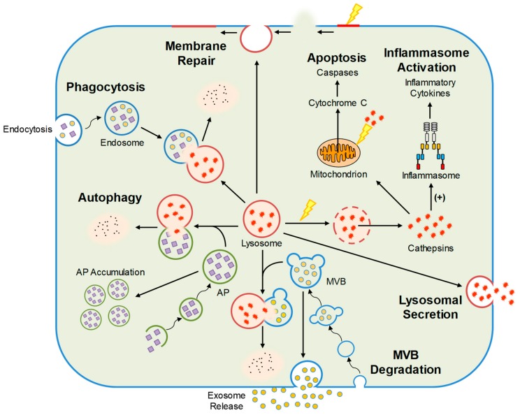
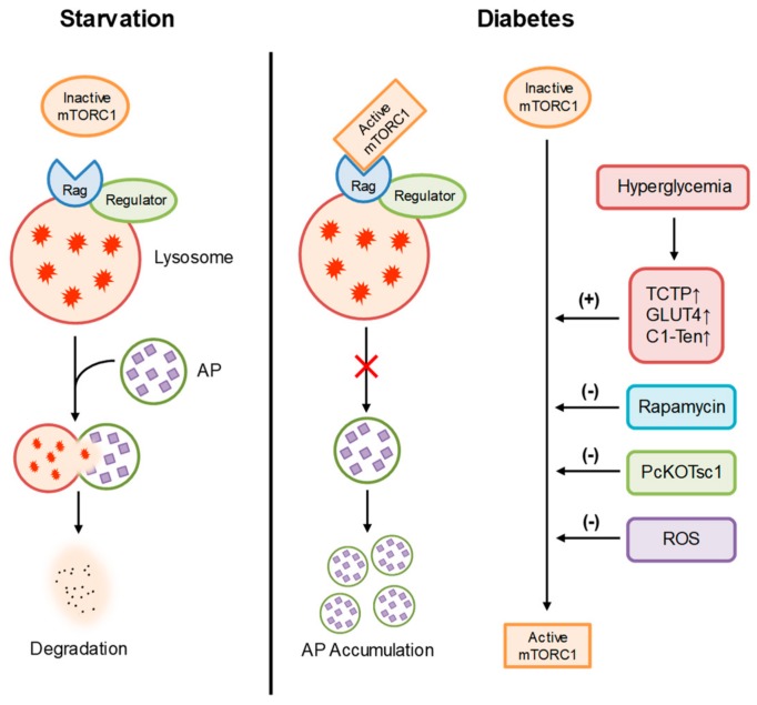
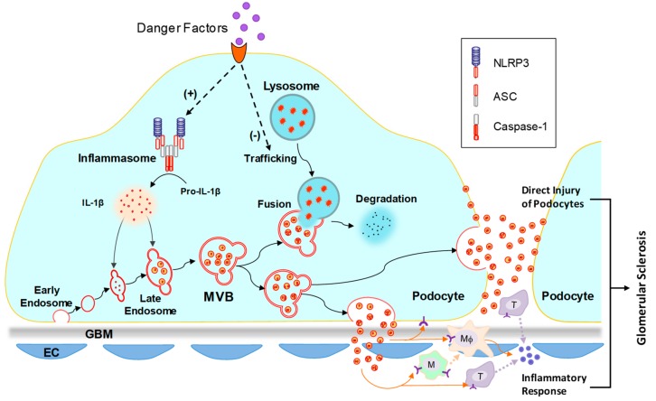
Podocytes are visceral epithelial cells covering the outer surface of glomerular capillaries in the kidney. Blood is filtered through the slit diaphragm of podocytes to form urine. The functional and structural integrity of podocytes is essential for the normal function of the kidney. As a membrane-bound organelle, lysosomes are responsible for the degradation of molecules via hydrolytic enzymes. In addition to its degradative properties, recent studies have revealed that lysosomes may serve as a platform mediating cellular signaling in different types of cells. In the last decade, increasing evidence has revealed that the normal function of the lysosome is important for the maintenance of podocyte homeostasis. Podocytes have no ability to proliferate under most pathological conditions; therefore, lysosome-dependent autophagic flux is critical for podocyte survival. In addition, new insights into the pathogenic role of lysosome and associated signaling in podocyte injury and chronic kidney disease have recently emerged. Targeting lysosomal functions or signaling pathways are considered potential therapeutic strategies for some chronic glomerular diseases. This review briefly summarizes current evidence demonstrating the regulation of lysosomal function and signaling mechanisms as well as the canonical and noncanonical roles of podocyte lysosome dysfunction in the development of chronic glomerular diseases and associated therapeutic strategies.

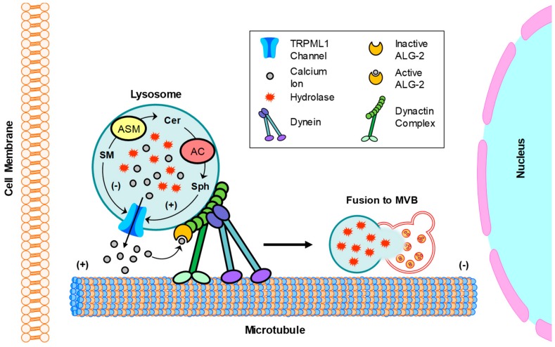
Keywords: chronic glomerular diseases; exosome; lysosome; podocyte; sphingolipids.


Conflict of interest statement

The authors declare no conflict of interest. The funders had no role in the design of the study; in the collection, analyses, or interpretation of data; in the writing of the manuscript, or in the decision to publish the results.

Figures

Figure 1
Figure 1
Cellular functions regulated by lysosomes including autophagy, MVB degradation, lysosomal secretion, phagocytosis, inflammasome activation, membrane repair, and apoptosis. AP, autophagosome; MVB, multivesicular body.
Figure 2
Figure 2
Regulation of mTORC1 in podocytes. During starvation, inactive mTORC1 has no effects on lysosome function and autophagic flux in podocytes. During diabetic nephropathy (DN), hyperglycemia upregulates TCTP, GLUT3, and C1-Ten, leading to overactivation of mTORC1 and inhibition of autophagy. As a specific mTORC1 inhibitor, rapamycin can prevent lysosome dysfunction, autophagic deficiency, and podocyte injury during diabetes. Reactive oxygen species (ROS) produced by mitochondria has also been reported to prevent mTORC1 activation in podocytes during diabetes.
Figure 3
Figure 3
Mechanism of inflammatory exosome release in podocytes. It has been reported that in podocytes, pathological stimuli may induce inflammasome activation and lysosome dysfunction. Activated inflammasome produces proinflammatory cytokines, such as IL-1β and IL-18. Lysosome dysfunction leads to reduced MVB degradation and increased exosome release. The proinflammatory cytokines in podocytes may be released through exosome release. The released inflammatory exosomes may induce direct injury of podocytes and inflammatory response, leading to the development of glomerular sclerosis. NLRP3, nucleotide-binding oligomerization domain-like receptor containing pyrin domain 3; ASC, adaptor molecule apoptosis-associated speck-like protein containing a caspase recruitment domain; IL-1β, interleukin-1β; GBM, glomerular basement membrane; EC, endothelial cell.
Figure 4
Figure 4
Regulation of lysosome trafficking by sphingolipids in podocytes. Dynein-mediated retrograde transport of lysosomes promotes their fusion with MVB. This transport is dependent on the TRPML1 channel-mediated Ca2+ release. In the lysosome, ASM converts SM into CER and AC converts CER to Sph. These sphingolipids, SM, Cer, and Sph had different effects on TRPML1 channel activity in podocytes, with inhibition by SM, no effect from Cer, but enhancement by Sph. SM, sphingomyelin; Cer; ceramide; Sph, sphingosine.

References

    1. Bainton D.F. The discovery of lysosomes. J. Cell Biol. 1981;91:66s–76s. doi: 10.1083/jcb.91.3.66s. - DOI - PMC - PubMed
    1. De Duve C. The lysosome turns fifty. Nat. Cell Biol. 2005;7:847–849. doi: 10.1038/ncb0905-847. - DOI - PubMed
    1. Essner E., Novikoff A.B. Localization of acid phosphatase activity in hepatic lysosomes by means of electron microscopy. J. Biophys Biochem. Cytol. 1961;9:773–784. doi: 10.1083/jcb.9.4.773. - DOI - PMC - PubMed
    1. Straus W. Isolation and biochemical properties of droplets from the cells of rat kidney. J. Biol. Chem. 1954;207:745–755. - PubMed
    1. Mundel P., Shankland S.J. Podocyte biology and response to injury. J. Am. Soc. Nephrol. 2002;13:3005–3015. doi: 10.1097/01.ASN.0000039661.06947.FD. - DOI - PubMed