. 2020 Feb 25;9(3):533.
doi: 10.3390/cells9030533.

Circumventing AKT-Associated Radioresistance in Oral Cancer by Novel Nanoparticle-Encapsulated Capivasertib

Affiliations

Circumventing AKT-Associated Radioresistance in Oral Cancer by Novel Nanoparticle-Encapsulated Capivasertib

Liwei Lang et al. Cells. .

Abstract

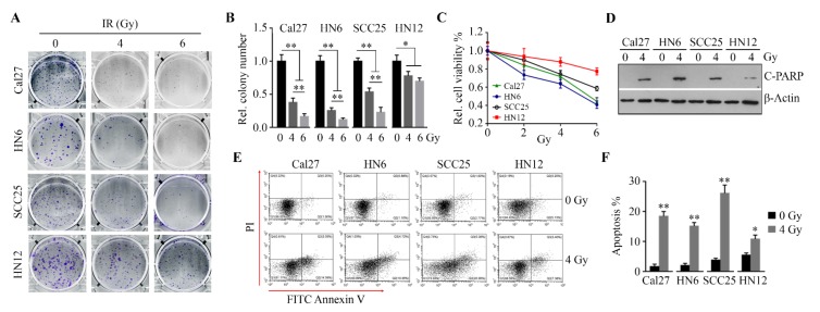
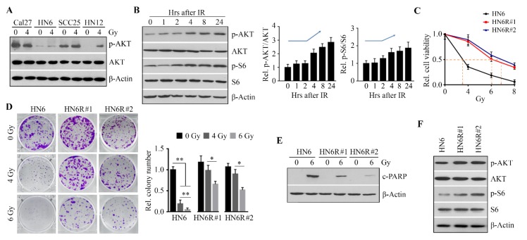
Background: Development of radioresistance in oral squamous cell carcinoma (OSCC) remains a significant problem in cancer treatment, contributing to the lack of improvement in survival trends in recent decades. Effective strategies to overcome radioresistance are necessary to improve the therapeutic outcomes of radiotherapy in OSCC patients.

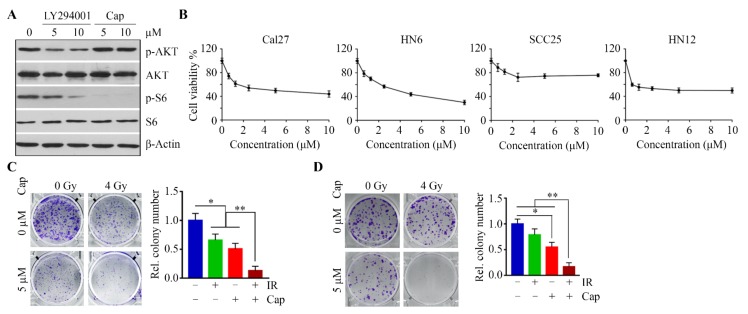
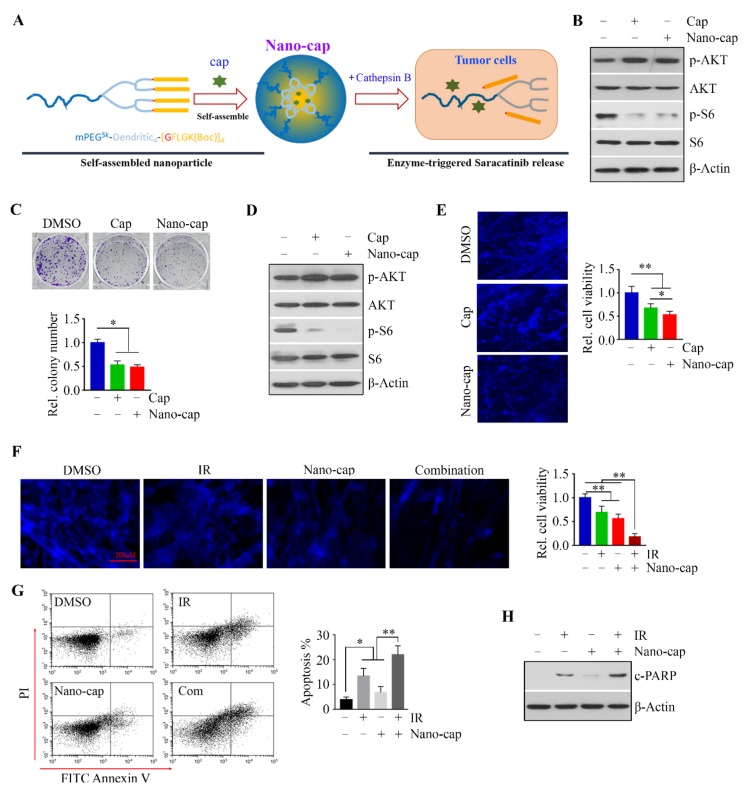
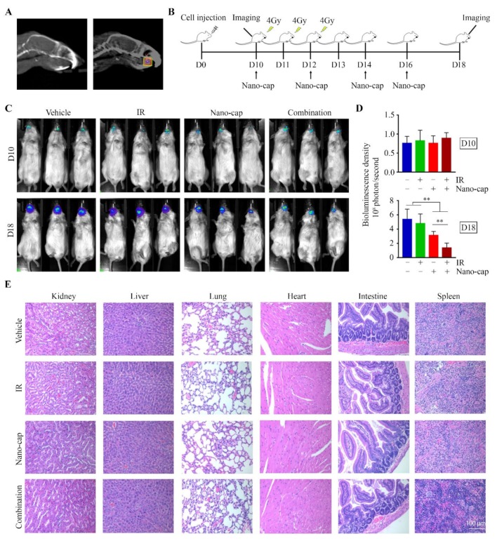
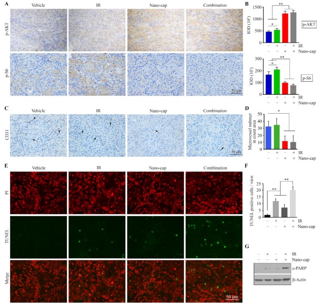
Methods: Cells and xenograft tumors were irradiated using the Small Animal Radiation Research Platform. AKT inhibitor capivasertib (AZD5363) was encapsulated into cathepsin B-responsible nanoparticles (NPs) for tumor-specific delivery. Cell viability was measured by alamarBlue, cell growth was determined by colony formation and 3D culture, and apoptosis was assessed by flow cytometry with the staining of Fluorescein isothiocyanate (FITC) Annexin V and PI. An orthotopic tongue tumor model was used to evaluate the in vivo therapeutic effects. The molecular changes induced by the treatments were assessed by Western blotting and immunohistochemistry.

Results: We show that upregulation of AKT signaling is the critical mechanism for radioresistance in OSCC cells, and AKT inactivation by a selective and potent AKT inhibitor capivasertib results in radiosensitivity. Moreover, relative to irradiation (IR) alone, IR combined with the delivery of capivasertib in association with tumor-seeking NPs greatly enhanced tumor cell repression in 3D cell cultures and OSCC tumor shrinkage in an orthotopic mouse model.

Conclusions: These data indicate that capivasertib is a potent agent that sensitizes radioresistant OSCC cells to IR and is a promising strategy to overcome failure of radiotherapy in OSCC patients.

Keywords: AKT/S6; OSCC; anticancer; capivasertib; nanoparticles; radioresistance.


Conflict of interest statement

The authors declare no conflict of interest.

Figures

Figure 1
Figure 1
Oral squamous cell carcinoma (OSCC) cells exhibit differential responses to irradiation (IR). (A, B) The effects of IR on the ability of OSCC cell lines to form colonies were determined on Day 14 after IR. The representative results and quantitative data from three independent experiments are shown in (A) and (B), respectively. (C) The effects of IR on OSCC cell viability were determined on Day 3 after IR. (D) The effect of IR on poly ADP-ribose polymerase (PARP) cleavage were determined in OSCC cell lines on Day 3 after IR. (E, F) The effects of IR on apoptosis were determined in OSCC cell lines using Fluorescein isothiocyanate (FITC) Annexin V Apoptosis Detection Kit with PI on Day 3 after IR. A representative result and quantitative data from three independent experiments are shown in (E) and (F), respectively. * p < 0.05; ** p < 0.01.
Figure 2
Figure 2
The upregulation of AKT/S6 signaling confers OSCC cells radioresistance. (A) AKT activity in OSCC cell lines one day after IR. (B) The levels of p-AKT and p-S6 in HN12 cells within 24 h following IR. A representative Western blotting and quantitative data from three independent experiments are shown in the left and right panels, respectively. (C) The effects of IR on viability of radioresistant HN6R#1 and HN6R#2 cells and radiosensitive parental HN6 cells were determined by alamarBlue on Day 5 after IR. (D) The effects of IR on the ability of radioresistant and radiosensitive HN6 cells to form colonies were measured on Day 14 after IR. A representative result and quantitative data from three independent experiments are shown in the left and right panels, respectively. (E) The effects of IR on PARP cleavage in radioresistant and radiosensitive HN6 cells were determined on Day 3 after IR. (F) p-AKT and p-S6 levels in radioresistant and radiosensitive HN6 cells on Day 1 after IR. * p < 0.05; ** p < 0.01.
Figure 3
Figure 3
Suppressing AKT/S6 signaling sensitizes OSCC cells to IR. (A) The effects of LY294002 and capivasertib (Cap) on S6 phosphorylation in HN12 cells on Day 1 after treatment. (B) The effects of capivasertib on viability of OSCC cell lines were determined on Day 3 after treatment. (C) The effects of combined capivasertib and IR treatment on the ability of HN12 cell to form colonies were determined on Day 14 after treatment. A representative result and quantitative data from three independent experiments are shown in the left and right panels, respectively. (D) The effect of combined capivasertib and IR treatment on the ability of HN6R cells to form colonies was determined on Day 14 after treatment. A representative result and quantitative data from three independent experiments are shown in the left and right panels, respectively. * p < 0.05; ** p < 0.01.
Figure 4
Figure 4
The addition of Nano-cap to IR greatly strengthens the cytotoxic effect on OSCC cells in a 3D SeedEZ scaffold. (A) Schematic representation of the self-assemble Nano-cap and its disassembly upon CTSB digestion. (B,D) The effects of Cap and Nano-cap on the p-AKT and p-S6 levels in HN12 cells were determined on Day 1 after treatment. Cells were seeded in a 2D culture dish (B) or SeedEZ scaffold (D). (C) The effects of Cap and Nano-cap on HN12 cell colony forming ability were determined on Day 14 after treatment. A representative result and quantitative data from three independent experiments are shown in the upper and lower panels, respectively. (E) The effects of Cap and Nano-cap on HN12 cell viability in 3D SeedEZ scaffold were determined on Day 7 after treatment. Representative images of 4′,6-diamidino-2-phenylindole (DAPI) staining and quantitative data of cell viability measured by alamarBlue are shown in the left and right panels, respectively (F) The effects of Nano-cap and IR alone or in combination on HN12 cell viability were determined on Day 7 after treatment. Representative images of DAPI staining and quantitative data of cell viability measured by alamarBlue are shown in the left and right panels, respectively. (G) The effects of Nano-cap and IR alone or in combination on HN12 cell apoptosis was determined on Day 3 after treatment. (H) The effects of Nano-cap and IR alone or in combination on PARP cleavage in HN12 cells were determined on Day 3 after treatment. * p < 0.05; ** p < 0.01.
Figure 5
Figure 5
Combined Nano-cap and IR treatment greatly enhances the negative effect on tumor growth relative to monotherapy in HN12-derived orthotopic xenograft mouse model. (A) CT images of the mouse head. Right panel, the square area inside the box indicates the location of the tumor xenograft in the tongue, which is the IR target. (B) The timeline of experimental procedures in vivo. HN12-derived tumor-bearing mice (n = 10/group) received IR and/or Nano-cap on day 10 (D10) after cell inoculation into tongue and were sacrificed on D18. (C) Representative bioluminescence images from the Xenogen IVIS-200 In Vivo imaging system showing the size of tongue tumors in mice at the beginning (D10) and the end (D18) of the indicated treatments. (D) Quantitative data (n = 10) of bioluminescence density of tongue tumors on D10 and D18. (E) Histology of major organs at the endpoint of each treatment. ** p < 0.01.
Figure 6
Figure 6
Nano-cap improves IR efficacy through suppressing AKT/S6 signaling and neovascularization in HN12-derived orthotopic xenograft mice. (A,B) p-AKT and p-S6 levels in tumor xenografts from mice receiving different treatments were determined by immunohistochemistry (IHC). Representative IHC images and quantification of IHC staining using Image pro-Plus6.0 are shown in (A,B), respectively. (C) CD31-postitive microvessels (arrows) in tumor xenografts from mice receiving different treatments were determined by IHC. (D) Quantification of the CD31-positive microvessels. (E,F) Apoptosis in tumor xenografts from mice receiving different treatments was determined by the Terminal deoxynucleotidyl transferase dUTP nick end labeling (TUNEL) assay. Representative TUNEL-stained xenograft sections are shown in (E) and quantification of TUNEL-positive tumor cells is shown in (F). (G) The effects of indicated treatments on PARP cleavage in the orthotopic xenograft model. * p < 0.05; ** p < 0.01.

References

    1. Chen W., Zheng R., Baade P.D., Zhang S., Zeng H., Bray F., Jemal A., Yu X.Q., He J. Cancer statistics in China, 2015. CA Cancer J. Clin. 2016;66:115–132. doi: 10.3322/caac.21338. - DOI - PubMed
    1. Siegiel R., Miller K., Jemal A. Cancer statistics, 2017. CA Cancer J. Clin. 2017;67:7–30. doi: 10.3322/caac.21387. - DOI - PubMed
    1. Bonner J.A., Harari P.M., Giralt J., Azarnia N., Shin D.M., Cohen R.B., Jones C.U., Sur R., Raben D., Jassem J. Radiotherapy plus cetuximab for squamous-cell carcinoma of the head and neck. N. Engl. J. Med. 2006;354:567–578. doi: 10.1056/NEJMoa053422. - DOI - PubMed
    1. Bussink J., van der Kogel A.J., Kaanders J.H. Activation of the PI3-K/AKT pathway and implications for radioresistance mechanisms in head and neck cancer. Lancet Oncol. 2008;9:288–296. doi: 10.1016/S1470-2045(08)70073-1. - DOI - PubMed
    1. Vivanco I., Sawyers C.L. The phosphatidylinositol 3-kinase–AKT pathway in human cancer. Nat. Rev. Cancer. 2002;2:489–501. doi: 10.1038/nrc839. - DOI - PubMed

Publication types