Review
. 2020 Mar 3;9(5):e015294.
doi: 10.1161/JAHA.119.015294. Epub 2020 Feb 28.

Apical Hypertrophic Cardiomyopathy: The Variant Less Known

Affiliations
Review

Apical Hypertrophic Cardiomyopathy: The Variant Less Known

Rebecca K Hughes et al. J Am Heart Assoc. .
No abstract available

Keywords: apical hypertrophic cardiomyopathy; cardiac magnetic resonance imaging; echocardiography; hypertrophic cardiomyopathy; imaging.


Figures

Figure 1
Figure 1
ECG and CMR in relative ApHCM. A, ECG demonstrates precordial T‐wave inversion and voltage criteria for LVH. B, Two‐chamber CMR demonstrates loss of apical tapering with relative but not absolute apical hypertrophy in diastole (Bi), systolic apical cavity obliteration (Bii) and LGE in the hypertrophied apex (Biii). ApHCM indicates apical hypertrophic cardiomyopathy; CMR, cardiovascular magnetic resonance; LGE, late gadolinium enhancement; LVH, left ventricular hypertrophy.
Figure 2
Figure 2
EKG in pure ApHCM. Voltage criteria for LVH and giant negative T‐wave inversion in precordial and inferolateral leads. ApHCM indicates apical hypertrophic cardiomyopathy; LVH, left ventricular hypertrophy.
Figure 3
Figure 3
CMR comparison of mixed ApHCM (A through C) and pure ApHCM (D through F), both with apical aneurysm formation. Long‐axis views of a patient with mixed ApHCM in diastole in 2‐chamber (Ai) and 4‐chamber (Aii), which in systole demonstrate midventricular obstruction but not total cavity obliteration due to persistence of apical chamber (Bi; Bii). The apical aneurysm contains LGE (Ci; Cii). A different patient with pure ApHCM has a thinned aneurysmal apex demonstrated in diastole on 2‐ (Di) and 4‐chamber views (Dii). In systole, the apical aneurysm becomes apparent (Ei; Eii) and contains LGE (Fi; Fii). ApHCM indicates apical hypertrophic cardiomyopathy; LGE, late gadolinium enhancement.
Figure 4
Figure 4
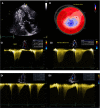
Transthoracic echocardiography in ApHCM. ApHCM with a small discrete apical chamber visible in the apical 3‐chamber view (A) and corresponding polar plot showing loss of longitudinal strain apically (B). At rest, continuous wave Doppler across the point of distal ventricular obstruction demonstrates a midsystolic peaking jet, followed by a drop in velocity prior to second peak representing paradoxical early diastolic jet flow, with gradients of 54 and 39 mm Hg, respectively (Ci). During Valsalva, systolic and diastolic jets merge, with a systolic intracavity gradient of 127 mm Hg, and a lengthening of the diastolic “tail” toward late diastole (Cii). By contrast, (D) demonstrates continuous wave Doppler traces from a patient with ApHCM and midcavity obstruction. At rest, there is midsystolic loss of Doppler alignment due to cavity obliteration, with corresponding Doppler dropout before paradoxical diastolic jet (Di). During Valsalva, the measured systolic gradient is unchanged, but the paradoxical diastolic jet gradient now exceeds 100 mm Hg with extension in duration to the end of diastole (Dii). ApHCM indicates apical hypertrophic cardiomyopathy.
Figure 5
Figure 5
Quantitative perfusion mapping in ApHCM. CMR pixelwise inline perfusion maps at rest (A), stress (B) in (i) basal, (ii) mid, (iii) apical short axis and (iv) 2‐chamber views in a patient with ApHCM and MVOCO. Stress perfusion defects are seen in the hypertrophied apex. Bull's‐eye plots are shown (rest C, stress D). There is 37% MBF reduction at stress (D) apically (1.47 mL/g per minute) vs 2.35 mL/g per minute in remote, non‐hypertrophied segments. Rest MBF(C) is 0.74 and 0.85 mL/g per minute, respectively. MPR is 1.99 in the apex and 2.76 in remote myocardium, indicating microvascular disease in the hypertrophied apex. Healthy volunteer stress MBF is 2 to 4 mL/g per minute. ApHCM indicates apical hypertrophic cardiomyopathy; CMR, cardiovascular magnetic resonance; MBF, myocardial blood flow; MPR, myocardial perfusion reserve; MVOCO, midventricular obstruction and cavity obliteration.

References

    1. Eriksson MJ, Sonnenberg B, Woo A, Rakowski P, Parker TG, Wigle ED, Rakowski H, Douglas Wigle E, Rakowski H, Toronto F. Long‐term outcome in patients with apical hypertrophic cardiomyopathy. J Am Coll Cardiol. 2002;39:638–645. - PubMed
    1. Sakamoto T, Tei C, Murayama M, Ichiyasu H, Hada Y. Giant T wave inversion as a manifestation of asymmetrical apical hypertrophy (AAH) of the left ventricle. Echocardiographic and ultrasono‐cardiotomographic study. Jpn Heart J. 1976;17:611–629. - PubMed
    1. Kubo T, Kitaoka H, Okawa M, Hirota THE. Clinical profiles of hypertrophic cardiomyopathy with apical phenotype. Circulation. 2009;73:2330–2336. - PubMed
    1. Klarich KW, Jost CHA, Binder J, Connolly HM, Scott CG, Freeman WK, Ackerman MJ, Nishimura RA, Tajik AJ, Ommen SR. Risk of death in long‐term follow‐up of patients with apical hypertrophic cardiomyopathy. Am J Cardiol. 2013;111:1784–1791. - PubMed
    1. Arad M, Penas‐Lado M, Monserrat L, Maron BJ, Sherrid M, Ho CY, Barr S, Karim A, Olson TM, Kamisago M, Seidman JG, Seidman CE. Gene mutations in apical hypertrophic cardiomyopathy. Circulation. 2005;112:2805–2811. - PubMed

Publication types

MeSH terms

LinkOut - more resources