. 2020 Apr 24;295(17):5795-5806.
doi: 10.1074/jbc.RA119.010697. Epub 2020 Feb 26.

Structural basis of cell-surface signaling by a conserved sigma regulator in Gram-negative bacteria

Affiliations

Structural basis of cell-surface signaling by a conserved sigma regulator in Gram-negative bacteria

Jaime L Jensen et al. J Biol Chem. .

Abstract

Cell-surface signaling (CSS) in Gram-negative bacteria involves highly conserved regulatory pathways that optimize gene expression by transducing extracellular environmental signals to the cytoplasm via inner-membrane sigma regulators. The molecular details of ferric siderophore-mediated activation of the iron import machinery through a sigma regulator are unclear. Here, we present the 1.56 Å resolution structure of the periplasmic complex of the C-terminal CSS domain (CCSSD) of PupR, the sigma regulator in the Pseudomonas capeferrum pseudobactin BN7/8 transport system, and the N-terminal signaling domain (NTSD) of PupB, an outer-membrane TonB-dependent transducer. The structure revealed that the CCSSD consists of two subdomains: a juxta-membrane subdomain, which has a novel all-β-fold, followed by a secretin/TonB, short N-terminal subdomain at the C terminus of the CCSSD, a previously unobserved topological arrangement of this domain. Using affinity pulldown assays, isothermal titration calorimetry, and thermal denaturation CD spectroscopy, we show that both subdomains are required for binding the NTSD with micromolar affinity and that NTSD binding improves CCSSD stability. Our findings prompt us to present a revised model of CSS wherein the CCSSD:NTSD complex forms prior to ferric-siderophore binding. Upon siderophore binding, conformational changes in the CCSSD enable regulated intramembrane proteolysis of the sigma regulator, ultimately resulting in transcriptional regulation.

Keywords: TonB-dependent transducer; X-ray crystallography; X-ray scattering; bacterial signal transduction; biophysics; cell surface signaling; inner membrane sigma regulator; metal homeostasis; protein structure; structural biology.


Conflict of interest statement

The authors declare that they have no conflicts of interest with the contents of this article

Figures

Figure 1.
Figure 1.
A, domain organization of PupR (an ASD, transmembrane region (TM), and CCSSD)) and PupB (a signal peptide (SP), NTSD, TonB box (region that interacts with the TonB complex), plug, β-barrel, and C-terminal TonB box)). Regions included in the expression constructs are colored. B, affinity pulldown assays to detect interaction of GST-tagged PupB NTSD and different MBP-tagged PupR CCSSD fragments as indicated. Equivalent aliquots of the clarified lysate from a co-expression of the two component proteins were applied to either amylose affinity agarose or GSH-Sepharose resins. Each resin was washed, then protein was eluted and analyzed by Coomassie-stained SDS-PAGE. The + sign above each lane indicates which resin was used for each experiment. The masses (kDa) of molecular weight markers are indicated in the first lane.
Figure 2.
Figure 2.
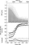
Global analysis of ITC isotherms for PupR CCSSD titrated into PupB NTSD. The heats of binding (top panel), the isotherms with the curves for the global model (middle panel), and residuals of the global model fit (bottom panel) for the triplicate experiments are shown in black, gray, and light gray.
Figure 3.
Figure 3.
CD spectra and melting curves. A, CD spectra of PupB NTSD (dashes), PupR CCSSD (dots), and the complex (solid); B, PupR CCSSD melting curve; C, PupB NTSD melting curve; and D, the complex melting curve. Unfolding (heating; black squares) and refolding (cooling; open circles) data points are shown. The Boltzmann fits to the melting curves are shown.
Figure 4.
Figure 4.
The X-ray crystal structure of the PupR CCSSD:PupB NTSD complex. Ribbon and transparent surface representations are colored purple for the PupR CCSSD and green for the PupB NTSD. The two CCSSD subdomains, the CJM and STN, are indicated.
Figure 5.
Figure 5.
Unique structural features of the PupR CCSSD. All structures are displayed in ribbon, rainbow color-ramped from blue at the N terminus to red at the C terminus. A, the CJM subdomain has a novel all-β-fold. B, the STN subdomain of the CCSSD is shown with the conserved residues L252, L259, L274, L266, L305, and F289 from the “LLLV” region in stick. C, the PupB NTSD, displayed in a superimposable orientation to the STN subdomain in B.
Figure 6.
Figure 6.
SEC-SAXS analysis of the CCSSD and CCSSD:NTSD complex. A, Guinier plot of the low q region. B, distance distribution P(r) for the experimental data (black lines), the theoretical curve calculated from the CCSSD crystal structure (purple line), and the CCSSD:NTSD complex (gray dashed line). Kratky plots of the (C) CCSSD and (D) the CCSSD:NTSD complex are shown. E, experimental scattering profile for the CCSSD, fit with the theoretical scattering profiles calculated from the rigid crystal structure of the CCSSD only (purple) and the flexible model derived from MultiFoxS, generated by structural conformation sampling (dark purple). F, experimental scattering profile for the complex, fit with the theoretical scattering profiles calculated from crystals structures of the CCSSD only (purple) and the CCSSD:NTSD complex (gray). χ values for each fit are indicated.
Figure 7.
Figure 7.
Affinity pulldown assays to detect interaction between different GST-tagged PupB NTSD and MBP-tagged PupR CCSSD mutants. Wild-type interaction between the PupR CCSSD and the PupB NTSD (second and third lanes). Residues stabilizing the PupR CCSSD:PupB NTSD interface were mutated as follows PupB Q69K (fourth and fifth lanes), PupB H72D (sixth and seventh lanes), PupB L74A (eighth and ninth lanes), PupR M251A (10th and 11th lanes), PupR S286A (12th and 13th lanes), and PupR T288A (14th and 15th lanes). The Coomassie-stained SDS-PAGE gel is shown. The + sign above each lane indicates which affinity resin was used for each experiment, as in Fig. 1. The masses of molecular weight markers are indicated in the first lane.
Figure 8.
Figure 8.
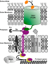
Schematic of the proposed CSS activation model. The proposed model starts with 1) the CSS system being primed by the TBDT NTSD:CCSSD sigma regulator interaction that stabilizes the sigma regulator; 2) ferric siderophore binding triggers signals for, 3), a and b, regulated intramembrane proteolysis, resulting in 4) release of the sigma regulator:sigma factor complex to activate transcription of iron import genes.

References

    1. Noinaj N., Guillier M., Barnard T. J., and Buchanan S. K. (2010) TonB-dependent transporters: regulation, structure, and function. Annu. Rev. Microbiol. 64, 43–60 10.1146/annurev.micro.112408.134247 - DOI - PMC - PubMed
    1. Llamas M. A., Imperi F., Visca P., and Lamont I. L. (2014) Cell-surface signaling in Pseudomonas: stress responses, iron transport, and pathogenicity. FEMS Microbiol. Rev. 38, 569–597 10.1111/1574-6976.12078 - DOI - PubMed
    1. Jensen J. L., Balbo A., Neau D. B., Chakravarthy S., Zhao H., Sinha S. C., and Colbert C. L. (2015) Mechanistic implications of the unique structural features and dimerization of the cytoplasmic domain of the Pseudomonas sigma regulator, PupR. Biochemistry 54, 5867–5877 10.1021/acs.biochem.5b00826 - DOI - PMC - PubMed
    1. Edgar R. J., Xu X., Shirley M., Konings A. F., Martin L. W., Ackerley D. F., and Lamont I. L. (2014) Interactions between an anti-sigma protein and two sigma factors that regulate the pyoverdine signaling pathway in Pseudomonas aeruginosa. BMC Microbiol. 14, 287 10.1186/s12866-014-0287-2 - DOI - PMC - PubMed
    1. Enz S., Mahren S., Stroeher U. H., and Braun V. (2000) Surface signaling in ferric citrate transport gene induction: interaction of the FecA, FecR, and FecI regulatory proteins. J. Bacteriol. 182, 637–646 10.1128/JB.182.3.637-646.2000 - DOI - PMC - PubMed

Publication types

Substances

Supplementary concepts

LinkOut - more resources