. 2020 Feb 27;11(1):1107.
doi: 10.1038/s41467-020-14446-2.

Mechanically and biologically skin-like elastomers for bio-integrated electronics

Affiliations

Mechanically and biologically skin-like elastomers for bio-integrated electronics

Shuo Chen et al. Nat Commun. .

Abstract

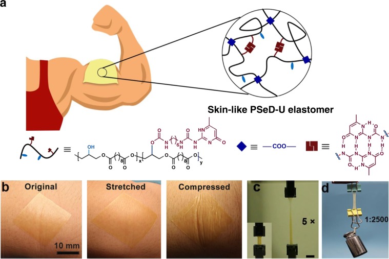
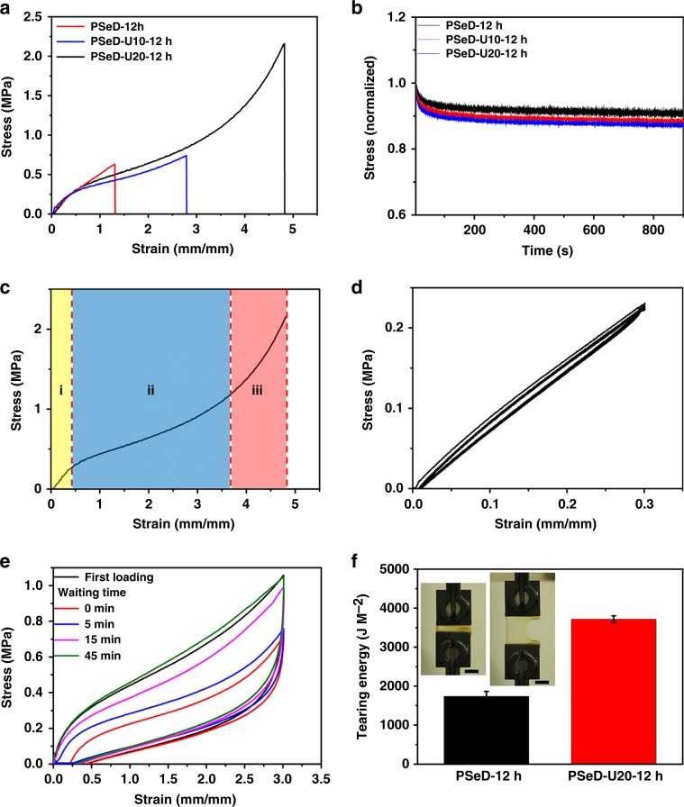
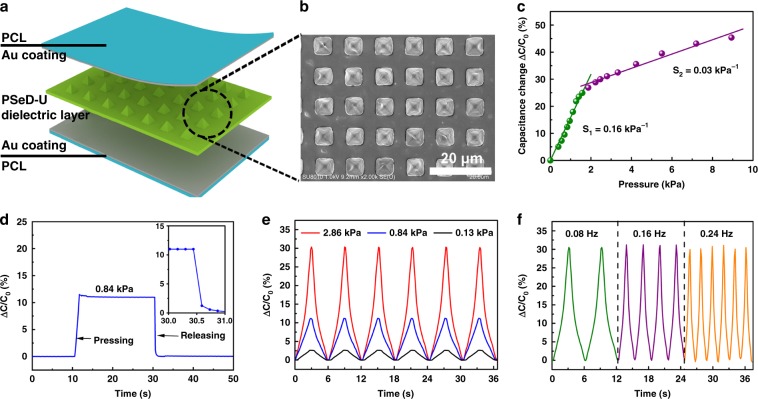
The bio-integrated electronics industry is booming and becoming more integrated with biological tissues. To successfully integrate with the soft tissues of the body (eg. skin), the material must possess many of the same properties including compliance, toughness, elasticity, and tear resistance. In this work, we prepare mechanically and biologically skin-like materials (PSeD-U elastomers) by designing a unique physical and covalent hybrid crosslinking structure. The introduction of an optimal amount of hydrogen bonds significantly strengthens the resultant elastomers with 11 times the toughness and 3 times the strength of covalent crosslinked PSeD elastomers, while maintaining a low modulus. Besides, the PSeD-U elastomers show nonlinear mechanical behavior similar to skins. Furthermore, PSeD-U elastomers demonstrate the cytocompatibility and biodegradability to achieve better integration with tissues. Finally, piezocapacitive pressure sensors are fabricated with high pressure sensitivity and rapid response to demonstrate the potential use of PSeD-U elastomers in bio-integrated electronics.


Conflict of interest statement

The authors declare no competing interests.

Figures

Fig. 1
Fig. 1. Schematic and performance of skin-like PSeD-U elastomers.
a The schematic illustration of the skin-like PSeD-U elastomers with physical and covalent hybrid crosslinked structures. b Photographs of spin-coated ultrathin PSeD-U20 film (70 µm thick) attached onto human skin: original, stretched, and compressed, illustrated its high conformability. Scale bar = 10 mm. c Photographs of original and five times stretched PSeD-U20-12h elastomer specimen showed the high stretchability of PSeD-U elastomers. Scale bar = 10 mm. d PSeD-U20-12h elastomer (40 mg) could hold a weight of 100 g (2500× its own weight) revealing its high strength and toughness.
Fig. 2
Fig. 2. Skin-like mechanical properties of PSeD-U elastomers.
a Typical tensile stress–strain curves of PSeD-U-12h elastomers with different densities of hydrogen bond while practically identical covalent crosslinking densities. b The stress relaxation curves of samples stretched at 5% strain for 900 s. c Dependence of stress on the strain, the stress–strain curve can be divided into three regions depending on the ranges of strain. d Cyclic tensile tests with a strain of 30%. e The tensile stress–strain curves of loading-unloading cycles with 300% strain with different waiting time between two consecutive loadings. f The comparison of tear energy of PSeD-12h and PSeD-U20-12h. The insert photograph is a notched PSeD-U20-12h specimen with a strain of 300%, showing its high tear resistance. Scale bar = 10 mm.
Fig. 3
Fig. 3. The degradation behaviors and cytocompatibility of PSeD-U elastomers.
a In vitro enzymatic degradation of PSeD and PSeD-U elastomers in lipase DPBS solutions (0.1 M) at 37 °C. b Tensile strength of PSeD-U20-12h at a strain of 200% after degradation for 0, 2, 4, 6, 8 h, showing a gradual decrease of mechanical strength. c Images of NIH 3T3 fibroblasts in live/dead assays of TCPS, PLGA, PSeD, and PSeD-U20. A large amount of live cells (green) and few dead cells (red) at all surface, indicating the good biocompatibility of PSeD-U (magnification = ×100; scale bar = 100 μm). d The MTT assay of NIH 3T3 fibroblasts cells cultured on TCPS, PLGA, PSeD, and PSeD-U20 elastomers. There was no significant difference between different materials at the same time point. Statistical significance compared with previous time point was marked as *p < 0.05 or **p < 0.01.
Fig. 4
Fig. 4. Schematic and characterization of PSeD-U elastomers based piezocapacitive pressure sensor.
a Schematic of the structure of the piezocapacitive pressure sensor. b SEM images of the microstructured PSeD-U20-12h elastomer dielectric film. c Pressure–response curve of the piezocapacitive pressure sensor. d Response times of the pressure sensor with the pressure of 0.84 kPa. The inset shows the response time of the pressure sensor to pressure release. e Dynamic responses of the pressure sensor at various pressures with a frequency of 0.08 Hz. f Dynamic responses of the pressure sensor at various frequencies with a pressure of 2.86 kPa.

References

    1. Hwang S-W, et al. A physically transient form of silicon electronics. Science. 2012;337:1640–1644. doi: 10.1126/science.1226325. - DOI - PMC - PubMed
    1. Kim D-H, et al. Epidermal electronics. Science. 2011;333:838–843. doi: 10.1126/science.1206157. - DOI - PubMed
    1. Wu W, Haick H. Materials and wearable devices for autonomous monitoring of physiological markers. Adv. Mater. 2018;30:e1705024. doi: 10.1002/adma.201705024. - DOI - PubMed
    1. Zhong J, et al. Stretchable self‐powered fiber‐based strain sensor. Adv. Funct. Mater. 2015;25:1798–1803. doi: 10.1002/adfm.201404087. - DOI
    1. Bauer S, et al. 25th anniversary article: a soft future: from robots and sensor skin to energy harvesters. Adv. Mater. 2014;26:149–162. doi: 10.1002/adma.201303349. - DOI - PMC - PubMed

Publication types

MeSH terms