. 2020 May;119(5):1583-1595.
doi: 10.1007/s00436-020-06629-z. Epub 2020 Feb 27.

Ultrastructure and cytochemistry of intrauterine embryonic and larval stages of Ityogonimus lorum (Digenea: Brachylaimidae) involving transitory development of ciliated miracidia

Affiliations

Ultrastructure and cytochemistry of intrauterine embryonic and larval stages of Ityogonimus lorum (Digenea: Brachylaimidae) involving transitory development of ciliated miracidia

Zdzisław Świderski et al. Parasitol Res. 2020 May.

Abstract

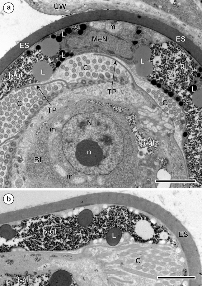
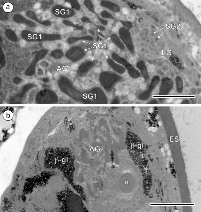
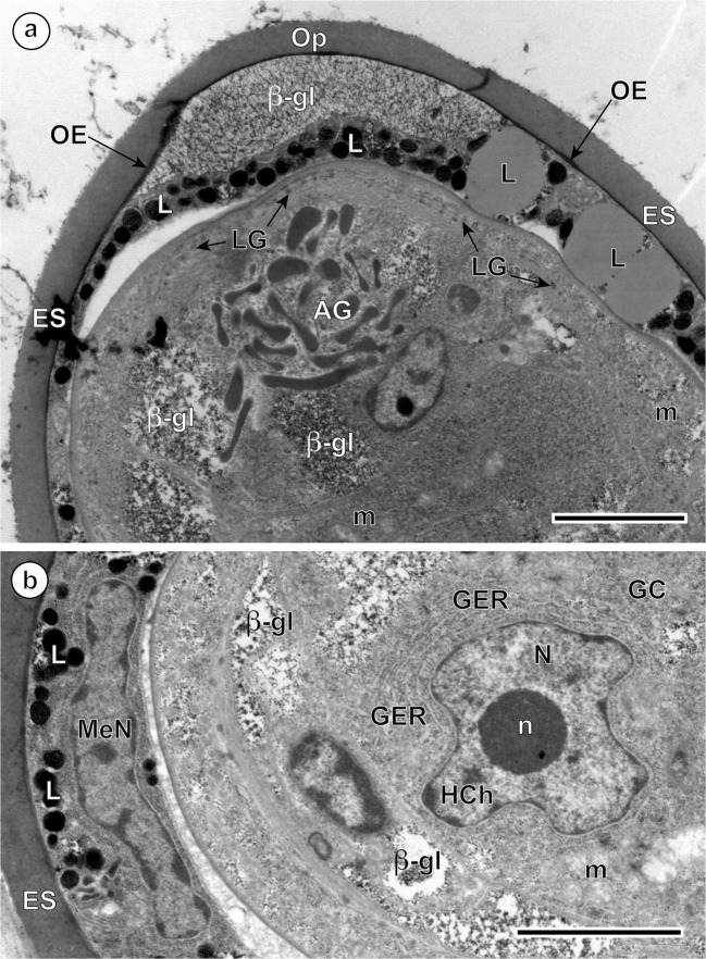
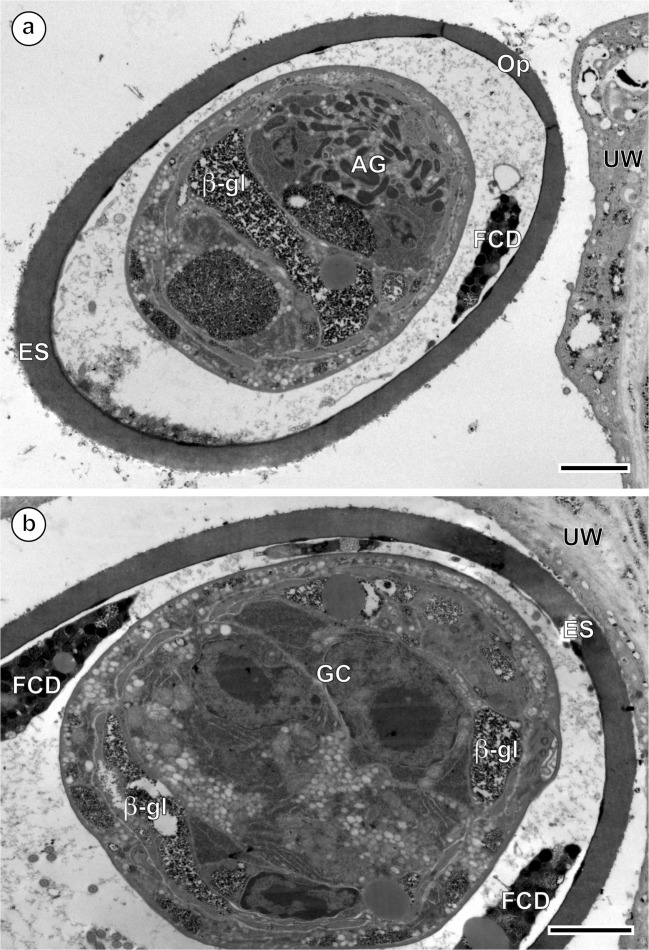
Results of the present study provide ultrastructural evidence that miracidial morphogenesis is fully completed within the intrauterine eggs while in the most posterior uterine regions of Ityogonimus lorum, a digenean parasite of an Iberian mole, Talpa occidentalis (Eulipotyphla, Talpidae). Using transmission electron microscopy (TEM), the ultrastructural characteristics of diverse cell types and their organelles of these developing embryos and fully formed miracidia within the eggshell were examined. The eggshell and embryonic envelopes are similar to those described previously by many authors for other digeneans. However, the developing miracidia are unique among previously described digeneans in possessing transitory cilia during larvigenesis, but completely lacking cilia in fully formed miracidium larvae. The evidence for completion of miracidial maturation in intrauterine eggs is based on the presence of the following structures: (1) transitional stage of ciliated differentiating miracidial epithelium; (2) apical and lateral glands, characteristic for digenean miracidia; and (3) fully developed germinative cells grouped together in the germinative sac localized in the posterior region of the miracidium. The protonephridial system with its characteristic flame cells and the nervous system with diverse types of neurons and nerve centers, which are characteristic for other digenean species reported until now, are absent from all these developmental stages of I. lorum. Based on these observations, we hypothesize that the life cycle of I. lorum is entirely terrestrial, involving passive transmission by ingestion of eggs containing unciliated miracidia to the first intermediate host.

Keywords: Brachylaimidae; Cytochemistry; Intrauterine embryogenesis; Ityogonimus lorum; Miracidial morphogenesis; Ultrastructure.


Conflict of interest statement

The authors declare that they have no conflict of interest.

Figures

Fig. 1
Fig. 1
Four consecutive stages of embryogenesis and larvigenesis in the intrauterine eggs of Ityogonimus lorum. We added shading and coloration to aid in interpreting important features of each layer. a A newly formed egg of I. lorum in the ootype region, composed of a fertilized ovum (Ov) and several vitellocytes (VC), all surrounded by a thin discontinuous layer of the differentiating eggshell (ES). b The early embryo composed of several micromeres (Mi) in the stage of forming two embryonic envelopes, the outer (OE) and inner (IE) envelopes. c Advanced ciliated stage of miracidial differentiation and maturation. Note the presence of ciliated tegument (CT), a single apical gland (AG), two lateral glands (LG), and numerous undifferentiated miracidial cells (MC). d Final stage of mature non-ciliated larvae. Note the presence of numerous somatic miracidial cells (SC) and several germinative cells (GC), grouped together in a sac-like germinative follicle (GS). In this stage, note also the complete autolysis of the ciliated tegument of the miracidium, entirely eliminated in the presence of several lysosome-like structures, appearing as areas of focal degradation (FCD). MaN, macromere nucleus; MeN, mesomere nucleus; Op, operculum
Fig. 2
Fig. 2
Ultrastructural details of Ityogonimus lorum egg envelopes. a Two large, concentric profiles of the granular endoplasmic reticulum (GER) in the cytoplasmic layer of the inner egg envelope (IE). The surface of embryonic tegument contains several vesicles of different sizes but is still unciliated. b A thick layer of the inner egg envelope (IE) cytoplasm with large, moderately saturated lipid droplets (L) and large accumulations of beta-glycogen particles (β-gl). The tegumental layer of the differentiating miracidium (Mir) shows presence of numerous ciliary rootlets (CR) and cilia (C), which are separated into individual zones by numerous tegumental processes (TP). ES, eggshell; OE, remnants of the outer envelope. Scale bars = 1 μm
Fig. 3
Fig. 3
Intrauterine eggs of Ityogonimus lorum illustrating part of the advanced ciliated stage of the miracidium in the advanced stage of differentiation and maturation. a Egg with the larval surface illustrating the transitory development of cilia on the tegumental larval surface, with zones of cilia (C) which are separated by zones of thin, elongated tegumental processes (TP). b Specific cytochemical localization of beta-glycogen particles (β-gl) in the inner egg envelope and in the somatic peripheral musculature of differentiating larvae after application of the test of Thiéry. Bl, blastomere; ES, eggshell; L, lipid droplets; m, mitochondrion; MaN, macromere nucleus; MeN, mesomere nucleus; n, nucleolus; N, nucleus; UW, uterine wall. Scale bars = 2 μm
Fig. 4
Fig. 4
Ultrastructural details of a progressive degeneration of miracidial cilia at one pole of the Ityogonimus lorum eggs. a Presence of the unciliated, smooth miracidial surface of the operculum-oriented pole of the majority of fully differentiated larvae. b Similar observations at higher magnification, which shows presence of only very limited number of cilia (C) at the pole of egg situated opposite the operculum. AG, apical gland; β-gl, beta-glycogen particles; ES, eggshell; L, lipid droplets; MeN, mesomere nucleus; OE, outer envelope; Op, operculum; UW, uterine wall. Scale bars (a) = 5 μm, (b) = 2 μm
Fig. 5
Fig. 5
Ultrastructural details of the two types of miracidial glands. a Micrograph showing differences between large, elongated secretory granules SG1 of the apical gland (AG) and spherical, much smaller and evidently less electron-dense granules SG2 of the lateral gland (LG). b Details of the perinuclear region of the apical gland after the test of Thiéry. Note a large nucleus of the apical gland with a prominent, spherical nucleolus (n) surrounded by a thick layer of granular cytoplasm containing several dense accumulations of beta-glycogen particles (β-gl). ES, eggshell. Scale bars (a) = 1 μm, (b) = 2 μm
Fig. 6
Fig. 6
Ultrastructural details of mature unciliated, smooth-surfaced eggs of Ityogonimus lorum. a Apical gland (AG) with its characteristic, elongated secretory granules. b A germinative cell (GC) with large irregularly shaped nucleus (N) and a prominent, electron-dense nucleolus (n). β-gl, beta-glycogen particles; ES, eggshell; L, lipid droplets; GER, granular endoplasmic reticulum; LG, lateral glands; m, mitochondria; MeN, mesomere nucleus; OE, outer envelope; Op, operculum. Scale bars = 2 μm
Fig. 7
Fig. 7
a, b Eggs of Ityogonimus lorum in the final stage of mature intrauterine unciliated miracidia. Note several focal cytoplasmic degradation (FCD) structures between the eggshell (ES) and unciliated miracidial tegument. AG, apical gland; GC, germinative cells; Op, operculum; UW, uterine wall. Scale bars = 2 μm
Fig. 8
Fig. 8
Diagram of hypothetical life cycle of Ityogonimus lorum, based on information available to date, including our data on functional ultrastructure of the miracidia

References

    1. Bruňanská M, Mackiewicz JS, Młocicki D, Świderski Z, Nebesářová J. Early intrauterine embryonic development in Khawia sinensis Hsü, 1935 (Cestoda, Caryophyllidea, Lytocestidae), an invasive tapeworm of carp (Cyprinus carpio): an ultrastructural study. Parasitol Res. 2012;110:1009–1017. doi: 10.1007/s00436-011-2590-2. - DOI - PubMed
    1. Butcher AR, Grove DI. Description of the life-cycle stages of Brachylaima cribbi n. sp. (Digenea: Brachylaimidae) derived from eggs recovered from human faeces in Australia. Syst Parasitol. 2001;49:211–221. doi: 10.1023/A:1010616920412. - DOI - PubMed
    1. Conn DB. Atlas of invertebrate reproduction and development. 2. New York: Wiley; 2000.
    1. Conn DB. Life cycles and biogeography of fish parasites: recent advances and future directions. Parassitologia. 2007;49(Suppl. 2):282.
    1. Conn DB. Invasion strategies of trematodes involving activity of the life-cycle stages between cercaria and metacercaria. In: Hodová I, Koubková B, editors. 18th helminthological days 2010: book of abstracts. Brno: MUNI Press, Masaryk University; 2010. pp. 22–23.