. 2020 Jul 2;56(1):1901965.
doi: 10.1183/13993003.01965-2019. Print 2020 Jul.

Quantitative analysis of airway obstruction in lymphangioleiomyomatosis

Affiliations

Quantitative analysis of airway obstruction in lymphangioleiomyomatosis

Stijn E Verleden et al. Eur Respir J. .

Abstract

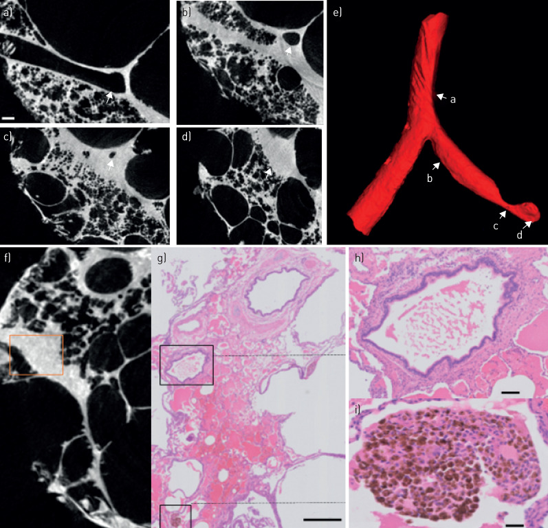
Lymphangioleiomyomatosis (LAM) is a rare, cystic lung disease with progressive pulmonary function loss caused by progressively proliferating LAM cells. The degree of airway obstruction has not been well investigated within the pathogenesis of LAM.Using a combination of ex vivo computed tomography (CT), microCT and histology, the site and nature of airway obstruction in LAM explant lungs was compared with matched control lungs (n=5 each). The total number of airways per generation, total airway counts, terminal bronchioles number and surface density were compared in LAM versus control.Ex vivo CT analysis demonstrated a reduced number of airways from generation 7 on (p<0.0001) in LAM compared with control, whereas whole-lung microCT analysis confirmed the three- to four-fold reduction in the number of airways. Specimen microCT analysis further demonstrated a four-fold decrease in the number of terminal bronchioles (p=0.0079) and a decreased surface density (p=0.0079). Serial microCT and histology images directly showed the loss of functional airways by collapse of airways on the cysts and filling of the airway by exudate.LAM lungs show a three- to four-fold decrease in the number of (small) airways, caused by cystic destruction which is the likely culprit for the progressive loss of pulmonary function.


Conflict of interest statement

Conflict of interest: S.E. Verleden has nothing to disclose. Conflict of interest: A. Vanstapel has nothing to disclose. Conflict of interest: L. De Sadeleer has nothing to disclose. Conflict of interest: B. Weynand has nothing to disclose. Conflict of interest: M. Boone has nothing to disclose. Conflict of interest: E. Verbeken has nothing to disclose. Conflict of interest: D. Piloni has nothing to disclose. Conflict of interest: D. Van Raemdonck has nothing to disclose. Conflict of interest: M. Ackermann has nothing to disclose. Conflict of interest: D.D. Jonigk has nothing to disclose. Conflict of interest: J. Verschakelen has nothing to disclose. Conflict of interest: W.A. Wuyts has nothing to disclose.

Figures

FIGURE 1
FIGURE 1
Comparison of lymphangioleiomyomatosis (LAM) and control lung tissue. a) Gross slice image of a LAM lung. b) Matched ex vivo computed tomography (CT) image in LAM. c) Representative subsample microCT image demonstrating evidence of cysts adjacent with limited normal surrounding tissue. d) Gross image of control lung. e) Ex vivo CT image of control lung at the same location. f) MicroCT image in control lung demonstrated normal lung tissue. a) and d) Scale bars=1 cm; c) and f) Scale bars: 1 mm.
FIGURE 2
FIGURE 2
Conventional computed tomography (CT)-based airway measures. a) The number of airways per generation is lower in lymphangioleiomyomatosis (LAM) compared to control from generation 7 till 10. b) The number of terminal bronchioles (TB) was decreased in LAM compared with control. c) Surface density is lower in LAM compared with control. d) Tissue percentage is decreased in LAM compared with control. e) Wall thickness of the terminal bronchioles in increased in LAM compared with control. **: p<0.01.
FIGURE 3
FIGURE 3
Whole lung micro-computed tomography (microCT) based airway measures. a) Complete airway segmentation of a right control lung. b) Airway segmentation of a right lymphangioleiomyomatosis (LAM) lung. c) The number of airways per generation based on whole lung microCT is lower in LAM compared with control. d) The number of airways per airway diameter. e) The proportion of airways relative to the total airway number per generation.
FIGURE 4
FIGURE 4
Serial subsample micro-computed tomography (microCT) images demonstrating collapse of airways on cystic structure. a) Mother branch. b) Bifurcation. c) Two separate airways. d) Airway lumen decrease in one airway (yellow), intact lumen in the other (white). e) Progressive luminal loss. f) Total luminal obliteration with an adjacent patent airway. g) 3D microCT illustration.
FIGURE 5
FIGURE 5
Comparison of serial micro-computed tomography (microCT) images (a–d) with matched haematoxylin-eosin (H&E) (e–h), Elastica van Gieson (EvG) (i–l), and HMB45 (m–t) stained serial sections. MicroCT imaging initially displays an open bronchus (a), where a bronchus is compressed (b) and where additional serial sectioning demonstrates further compression of the lumen (c-d). The corresponding H&E and EvG stains confirm the gradual complete compression of the bronchial lumen by cyst formation (arrows in f and h), as evidenced by preservation of the bronchial epithelium between these cysts. In addition, there are signs of severe haemorrhage, with abundant presence of red blood cells and haemosiderin-laden macrophages. The corresponding HMB45 images show a preserved airway wall in a patent bronchus (m and q), but abundant presence and infiltration of HMB45+ lymphangioleiomyomatosis (LAM) cells (magenta stained cells) in the walls of the compressed airways (r–t), potentially contributing to the airway obliteration with even complete loss of the airway lumen (t). Awy: airway. Scale bars: (a–d) 500 μm; (q–t) 100 μm.
FIGURE 6
FIGURE 6
Serial subsample micro-computed tomography (microCT) images demonstrating airway loss due to fibrosis. a) Mother branch. b) Airway with peribronchiolar fibrosis. c) Luminal narrowing. d) Luminal loss in region of fibrosis. e) 3D reconstruction demonstrating airway obstruction. f) MicroCT image showing a patent and obstructed bronchiole (highlighted with the rectangle). g) Corresponding haematoxylin-eosin (H&E) image showing filling of the airway with exudate. h) Higher magnification image showing filling of the airway. i) Evidence of haemosiderin-filled macrophages in close proximity of the airway. Scale bars: (g) 800 μm; (h) 100 μm; (i) 50 μm.
FIGURE 7
FIGURE 7
Structural morphology and vascularity of lymphangioleiomyomatosis (LAM). In comparison with the intact alveolar architecture of healthy control lungs (a), scanning electron micrographs of pulmonary LAM revealed large cystic honeycombing with (c, d) cystic walls showed intermingled plumb, spindle-shaped cell proliferation (blue arrows) whereas the inner cystic wall is largely covered by type II pneumocytes (red arrowheads). e) Microvascular corrosion casting depicts the thin-walled alveolar plexus of healthy control tissue. f) Microvascular architecture in pulmonary LAM demonstrated a distinct aberrant vascularity with sinusoid-like vessel and missing hierarchy. Scale bars: (a and b) 100 µm; (c–f) 20 µm.

Comment in

References

    1. McCormack FX. Lymphangioleiomyomatosis: a clinical update. Chest 2008; 133: 507–516. doi:10.1378/chest.07-0898 - DOI - PubMed
    1. Henske EP, McCormack FX. Lymphangioleiomyomatosis - a wolf in sheep's clothing. J Clin Invest 2012; 122: 3807–3816. doi:10.1172/JCI58709 - DOI - PMC - PubMed
    1. Baldi BG, Araujo MS, Freitas CSG, et al. . Evaluation of the extent of pulmonary cysts and their association with functional variables and serum markers in lymphangioleiomyomatosis (LAM). Lung 2014; 192: 967–974. doi:10.1007/s00408-014-9641-2 - DOI - PubMed
    1. Burger CD, Hyatt RE, Staats BA. Pulmonary mechanics in lymphangioleiomyomatosis. Am Rev Respir Dis 1991; 143: 1030–1033. doi:10.1164/ajrccm/143.5_Pt_1.1030 - DOI - PubMed
    1. Sobonya RE, Quan SF, Fleishman JS. Pulmonary lymphangioleiomyomatosis: quantitative analysis of lesions producing airflow limitation. Hum Pathol 1985; 16: 1122–1128. doi:10.1016/S0046-8177(85)80180-5 - DOI - PubMed

Publication types

MeSH terms