. 2020 Feb 27;10(1):3581.
doi: 10.1038/s41598-020-60333-7.

Fabrication of sharp silicon arrays to wound Caenorhabditis elegans

Affiliations

Fabrication of sharp silicon arrays to wound Caenorhabditis elegans

Jérôme Belougne et al. Sci Rep. .

Abstract

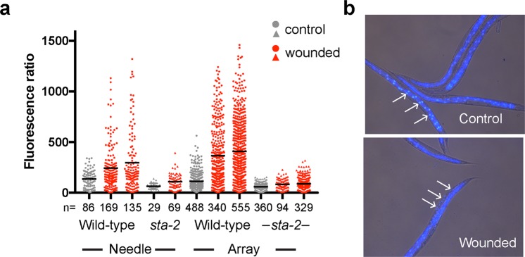
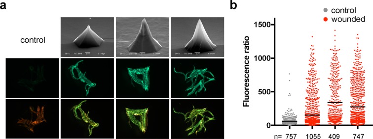
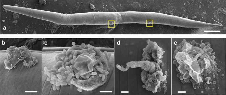
Understanding how animals respond to injury and how wounds heal remains a challenge. These questions can be addressed using genetically tractable animals, including the nematode Caenorhabditis elegans. Given its small size, the current methods for inflicting wounds in a controlled manner are demanding. To facilitate and accelerate the procedure, we fabricated regular arrays of pyramidal features ("pins") sharp enough to pierce the tough nematode cuticle. The pyramids were made from monocrystalline silicon wafers that were micro-structured using optical lithography and alkaline wet etching. The fabrication protocol and the geometry of the pins, determined by electron microscopy, are described in detail. We also used electron microscopy to characterize the different types of injury caused by these pins. Upon wounding, C. elegans expresses genes encoding antimicrobial peptides. A comparison of the induction of antimicrobial peptide gene expression using traditional needles and the pin arrays demonstrates the utility of this new method.


Conflict of interest statement

The authors declare no competing financial interests.

Figures

Figure 1
Figure 1
Drawing of a sample holder used for homogeneous KOH etching of multiple one-inch wafers (Autodesk 123 Design software). (a) Main support with magnets; (b) Arms for easy removal from etching solution; (c) One-inch (2.54 cm) diameter wafer to be etched.
Figure 2
Figure 2
Silicon pyramids in a 75 μm array etched in fresh 45 % KOH solution at T=63 °C, sample holder rotation speed 170 r.p.m for 100 minutes (a), 110 minutes (b), 114 minutes, followed by an additional 4 minutes, after removal and inspection with a stereomicroscope (c). The 3 images are at the same magnification. (d) Large array of homogeneous pins at the end of the etching period. Of the 342 pins in this electron photomicrograph, 130 retain the "hat” that is subsequently removed in a pure water ultrasound bath. Manufacturing parameters: 75 μm array; 750 ml fresh 45 % KOH; T=72 °C; 100 rpm. All the samples were tilted in the SEM at 70°.
Figure 3
Figure 3
Efficient wounding of C. elegans with a silicon array. (a) Quantification of the change of reporter gene expression provoked by wounding with a needle (5 left-hand columns), or arrays, worms carrying frIs7 in the wild-type or sta-2 mutant background (circles and triangles, respectively). The fluorescence ratio (Green/TOF in arbitrary but constant units) is shown for the indicated number of worms for each condition (n). The bars indicate the means. (b) Representative images of control (top) and wounded (bottom) adult wild-type worms stained with Hoechst 33258. In the control animals, only staining of the intestinal nuclei is apparent (arrows), while in the array-wounded worms nuclei in the epidermis (arrows) are also stained.
Figure 4
Figure 4
Wounding of C. elegans with different silicon arrays. (a) Top row shows SEM images of 3 different arrays (left to right: H75-42, H100-9, H75-46). Note that, as indicated, the magnification is not the same for all 3, hence the scale bars are different too. The other 2 rows are GFP (middle row) or mixed GFP/dsRed fluorescence signals for representative groups of worms carrying frIs7 picked because they appeared to have a normal morphology (i.e. were not badly wounded) after treatment with the indicated array, compared to non-wounded controls (left-hand column). (b) Quantification of the change of reporter gene expression provoked by wounding with the 3 arrays, in the same order as in a, in worms carrying frIs7. Details are as in the previous figure.
Figure 5
Figure 5
Wounds inflicted on C. elegans by silicon arrays. (a) SEM images of an adult hermaphrodite worm that showed normal movement and overall morphology and had strong nlp-29p::GFP reporter gene expression 6 hours after wounding. The worm has at least 2 lesions (boxed in yellow). These are shown at higher magnification in (b,c). In common with many lesions, the extruded material was found covered in bacteria. Two further wounds from a different worm are also shown (d,e). Scale bars, 100, 5, 2, 1, 1 micron, (a-e).
Figure 6
Figure 6
Artificial montage to illustrate the relative sizes of C. elegans and pins, with adult hermaphrodite worms fixed for EM placed directly on silicon pyramids in 100 μm (a) and 75 μm (b) arrays. Scale bars, 50 and 20 micron, (a,b).

References

    1. Pujol N, et al. Distinct innate immune responses to infection and wounding in the C. elegans epidermis. Curr. Biol. 2008;18:481–9. doi: 10.1016/j.cub.2008.02.079. - DOI - PMC - PubMed
    1. Hashmi S, et al. Genetic transformation of nematodes using arrays of micromechanical piercing structures. Bio. Techniques. 1995;19:766–770. - PubMed
    1. Trimmer, W. et al. Injection of DNA into plant and animal tissues with micromechanical piercing structures.In Proceedings IEEE Micro Electro Mechanical Systems. 1995, 111, 10.1109/MEMSYS.1995.472544 (IEEE, Amsterdam, Netherlands, 1995).
    1. Hopcroft MA, Nix WD, Kenny TW. What is the Young’s modulus of silicon? J. Microelectromechanical Syst. 2010;19:229–238. doi: 10.1109/JMEMS.2009.2039697. - DOI
    1. Fruhauf, J. Shape and Functional Elements of the Bulk Silicon Microtechnique (Springer-Verlag,Berlin/Heidelberg, 2005).

Publication types

MeSH terms