Case Reports
. 2020 Feb 27;15(2):e0229718.
doi: 10.1371/journal.pone.0229718. eCollection 2020.

Requirement of the acyl-CoA carrier ACBD6 in myristoylation of proteins: Activation by ligand binding and protein interaction

Affiliations
Case Reports

Requirement of the acyl-CoA carrier ACBD6 in myristoylation of proteins: Activation by ligand binding and protein interaction

Eric Soupene et al. PLoS One. .

Erratum in

Abstract

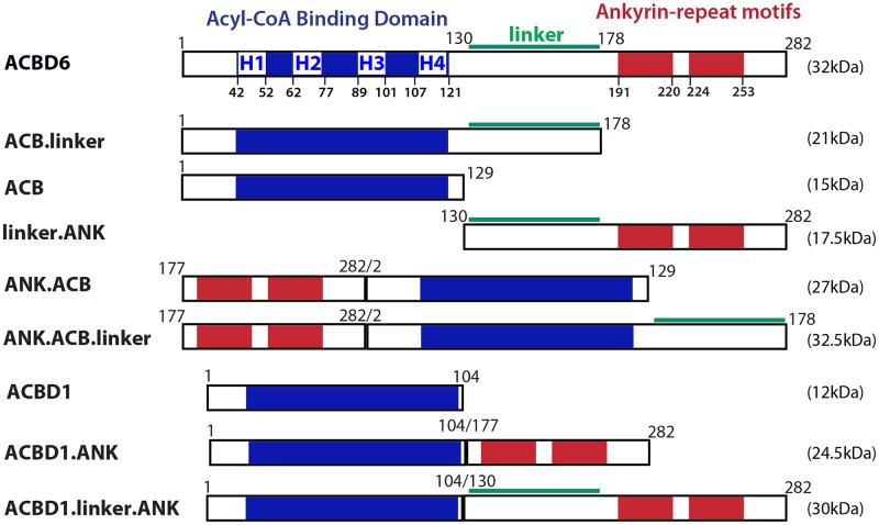
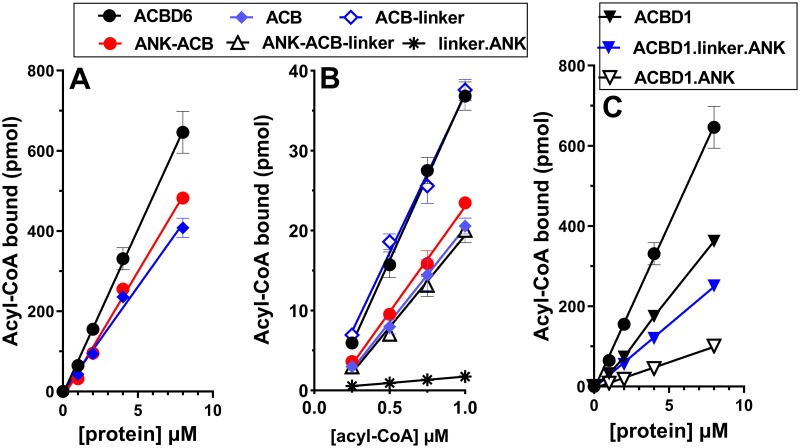
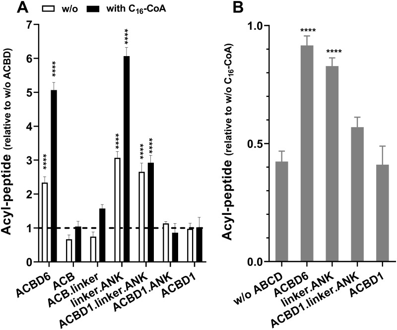
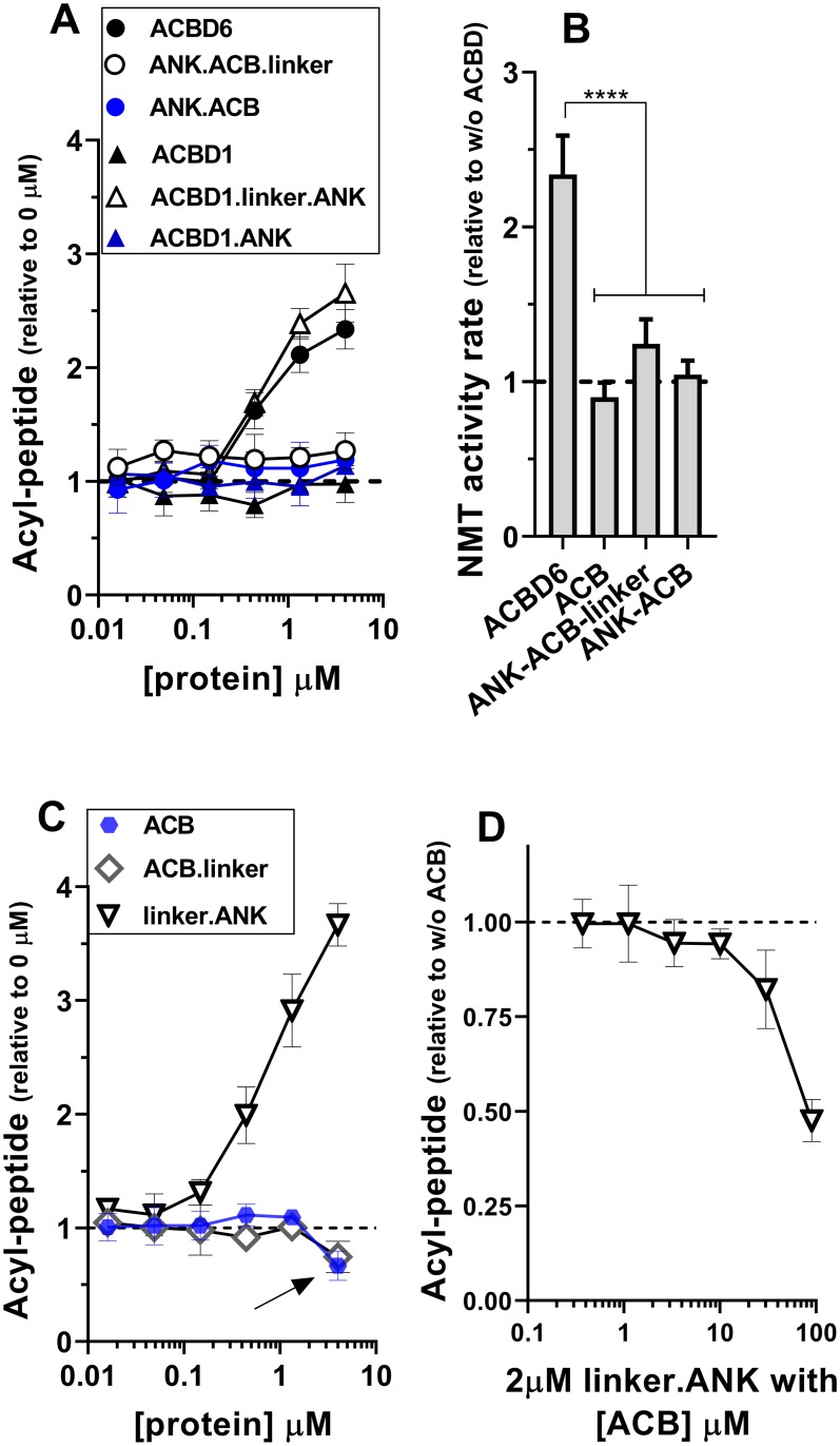
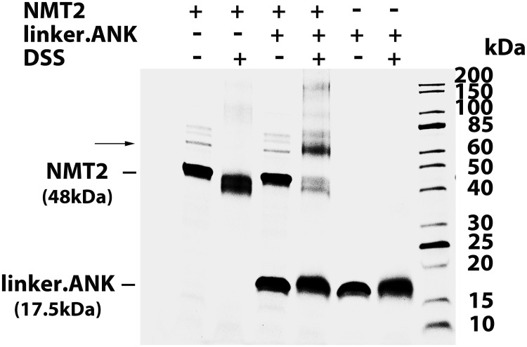
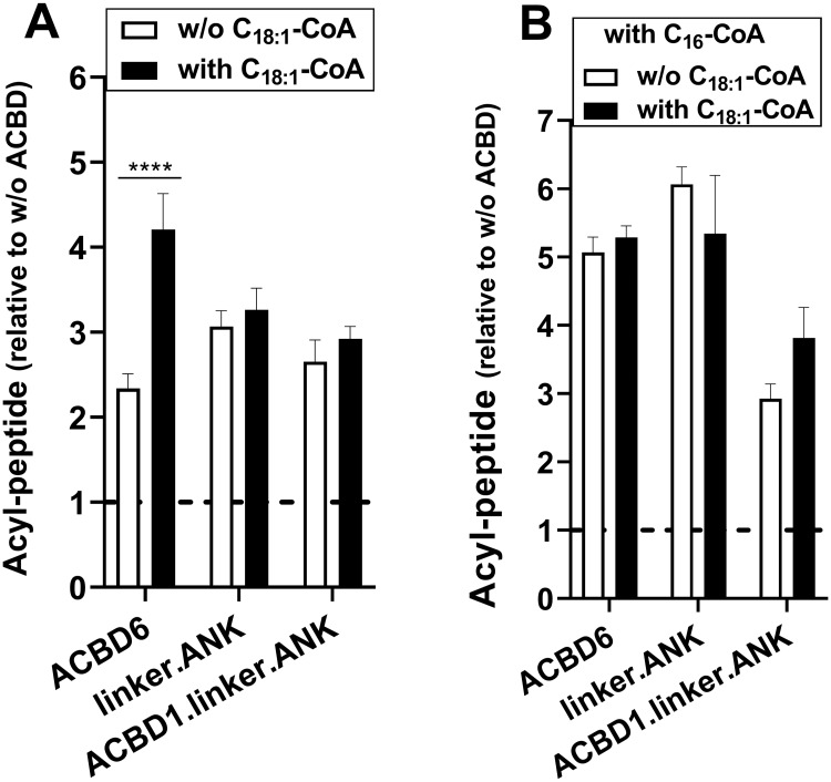
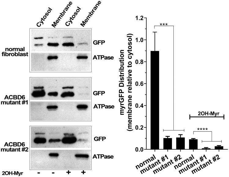
Glycine N-myristoylation is an essential acylation modification modulating the functions, stability, and membrane association of diverse cytosolic proteins in human cells. Myristoyl-CoA is the 14-carbon acyl donor of the acyltransferase reaction. Acyl-CoAs of a chain length compatible with the binding site of the N-myristoyltransferase enzymes (NMT) are competitive inhibitors, and the mechanism protecting these enzymes from unwanted acyl-CoA species requires the acyl-CoA binding protein ACBD6. The acyl-CoA binding domain (ACB) and the ankyrin-repeat motifs (ANK) of ACBD6 can perform their functions independently. Interaction of ANK with human NMT2 was necessary and sufficient to provide protection. Fusion of the ANK module to the acyl-CoA binding protein ACBD1 was sufficient to confer the NMT-stimulatory property of ACBD6 to the chimera. The ACB domain is dispensable and sequestration of the competitor was not the basis for NMT2 protection. Acyl-CoAs bound to ACB modulate the function of the ANK module and act as positive effector of the allosteric activation of the enzyme. The functional relevance of homozygous mutations in ACBD6 gene, which have not been associated with a disease so far, is presented. Skin-derived fibroblasts of two unrelated individuals with neurodevelopmental disorder and carrying loss of function mutations in the ACBD6 gene were deficient in protein N-myristoylation. These cells were sensitive to substrate analog competing for myristoyl-CoA binding to NMT. These findings account for the requirement of an ANK-containing acyl-CoA binding protein in the cellular mechanism protecting the NMT enzymes and establish that in human cells, ACBD6 supports the N-myristoylation of proteins.


Conflict of interest statement

The authors have declared that no competing interests exist.

Figures

Fig 1
Fig 1. Cartoon representation of the constructs used in this study.
The 4 α-helix (H1 to H4) of the acyl-CoA binding domain (blue), of the linker region (green), and of the two ankyrin-repeat motifs (red) of human ACBD6 (282 residues) are shown. For clarity, the different constructs are designed by the presence of the 3 regions: ACB domain (residues 1 to 129), linker (130 to 178), and ANK motifs (177 to 282). All constructs were produced with a hexahistidine tag at their carboxyl-terminal end and the calculated mass (kDa) of each protein is shown on the right. ACBD1 refers to isoform 1 of the product of the DBI gene.
Fig 2
Fig 2. Analysis of module deletion and switch on acyl-CoA binding function.
Binding assays were performed with 15μM 14C-C18:1-CoA and increasing concentrations of the indicated proteins (0 to 8μM) (Panel A and C) and with 2μM of the indicated proteins with increasing concentrations of 14C-C18:1-CoA (0.25 to 1μM) (Panel B). Error bars represent the standard deviations of values obtained from four measurements.
Fig 3
Fig 3. The C-terminal module is sufficient for NMT2 stimulation and protection.
Formation of the myristoyl-peptide by NMT2 was measured in the presence of the indicated proteins over a concentration range of 0.016 to 4μM for 3 min at 37°C. The full data set is presented in S1 Fig. The bar graphs summarize the values obtained at 4μM. Human NMT2 enzyme was added at a concentration of 50nM in the presence of 5μM C14-CoA and in the absence (white bars) or presence of excess competitor C16-CoA (50μM) (filled bars). Control reactions were performed in the absence of the ACBD constructs and of C16-CoA. The data are presented relative to the value obtained in the absence of ACBD (panel A) or in the absence of C16-CoA (panel B). Error bars represent the standard deviations of values obtained from three reactions. **** indicate a p value of <0.0001.
Fig 4
Fig 4. Role of the ACB domain and linker region in NMT2 activity.
Formation of the myristoyl-peptide by NMT2 was measured in the presence of the indicated proteins over a concentration range of 0.016 to 4μM (panel A and C) and with 2μM of the linker.ANK form in the presence of increasing concentration of the ACB form (0.4 to 90μM) (panel D). Activity of NMT2 was measured in the presence of 4μM of the indicated proteins from 0 to 8 min and the rates of formation of the acyl-peptide are reported relative to the values obtained in their absence (panel B). Control reactions were performed in the absence of the ACBD constructs (panel A, C) and in the absence of ACB (panel D). The data are presented relative to the values obtained in their absence. The decrease in acyl-peptide formation in the presence of the ACB and ACB.linker forms observed at 4μM is indicated with an arrow in panel C. Human NMT2 enzyme was added at a concentration of 50nM in the presence of 5μM C14-CoA. Error bars represent the standard deviations of values obtained from three reactions. **** indicate a p value of <0.0001.
Fig 5
Fig 5. The C-terminal module is sufficient for NMT2 interaction.
NMT2 (10μM) and the linker.ANK (30μM) recombinant protein were dialyzed together at 4°C for 16 hours. As indicated, the proteins were then reacted with the DSS reagent (1.2mM) for 2 hours at 4°C. Reactions were stopped by quenching with a solution of 100mM Tris-HCl pH 7.5. Proteins were separated on denaturing SDS-gradient gel (Any Kd, Bio-Rad). After electrophoresis, gel was stained with Gelcode Blue (Fisher Scientific). The panel shows the image of the gel with the position of NMT2 (48kDa), of linker.ANK (17.5kDa), and of a complex formed after treatment by DSS indicated on the left. The molecular masses of the ladder (Unstained Protein Standard, Broad Range; NewEnglandBiolabs) are shown on the right.
Fig 6
Fig 6. Acyl-CoA binding to the ACB domain enhances NMT2 activity.
Formation of the myristoyl-peptide by NMT2 (50nM) was measured in the presence of the indicated proteins (4μM) and with 5μM C14-CoA. As indicated, C16-CoA (50μM) and C18:1-CoA (20μM) were added in the reactions. Control reactions were performed in the absence of the ACBD constructs and the data are presented relative to the values obtained in their absence. Error bars represent the standard deviations of values obtained from three reactions. **** indicate a p value of <0.0001. Differences detected with the linker.ANK and ACBD1.linker.ANK forms did not reach statistical significance of p ≤0.05.
Fig 7
Fig 7. Spliced isoforms identification of human ACBD6-deficient fibroblasts.
Panel A. The usage of an alternative splice site in exon 6 (mutant #1) and the 5 bases deletion in exon 5 (mutant #2) are indicated as red boxes on the cartoon representation of ACBD6. The exon organization of the linker and the ANK-repeat motifs (ANK1 and ANK2) region is also shown. The positions of the two primer pairs (1/1’ and 2/2’) used for reversed transcription of ACBD6 mRNA are indicated. Panel B. Three cDNAs were identified with the RNA isolated from mutant #1 and are indicated as isoform a, b, and c. Separation of the cDNAs on 1.2% agarose gel is shown in the inset. Isoform a lacks exon 6 encoding the ANK1 motif; isoform b represents the splicing of exon 5 to the alternative splice acceptor site identified in exon 6, and result in the early translational stop removing the ANK1 and ANK2 motifs; isoform c is similar to isoform b but with an insertion of a sequence present in intron 5, and resulting in the disruption of ANK1. Because of the low level of expression of ACBD6 mRNA in those cells, the very low abundant isoform c might represent an immature pre-RNA of isoform b detected by end-point RT-PCR. Panel C. The five bases deletion affecting exon 5 of ACBD6 in mutant #2 is highlighted in red. The single isoform detected in these cells will produce a form truncated of the linker and ANK motifs. Panel D. Expression of ACBD6, NMT1 and NMT2 were determined by qRT-PCR, using ACTB as the reference mRNA. The values obtained with RNA isolated from the two mutant cells are reported relative to the values obtained with normal fibroblast. Note the logarithmic scale of the x axis. Error bars represent the standard deviations of values obtained from 4 measurements.
Fig 8
Fig 8. Decreased protection of NMT in ACBD6-deficient cells.
Fibroblasts were grown in 96-well plates and exposed to the indicated concentration of 2-hydroxymyristate (2-OH Myr) and myristate. At the indicated times, the medium was removed and cells were fixed in 10% ice-cold TCA, stained with the SRB dye and absorbance was read at 560nm. In panel B, values obtained in the presence of the drugs are reported relative to the values obtained in their absence. Error bars represent the standard deviations of values obtained from 4 measurements.
Fig 9
Fig 9. Decreased membrane localization of myrGFP in ACBD6-deficient cells.
Fibroblasts were transfected with a vector expressing a myrGFP construct and grown for 48 hours in the absence or the presence of 100μM 2-OH Myr. The mRNA levels of NMT1 and NMT2 were not affected by such treatment and were similar to those of untreated cells. Cells were harvested, lyzed and the cytosolic and membranes fractions were isolated. Proteins were separated on denaturing SDS-PAGE, transferred to PVDF membranes, and GFP and the membrane marker Na+/K+-ATPase α1 were detected with mouse monoclonal antibodies. The bar graph shows the quantification of the signals of GFP in the membranes reported relative to the values obtained with the cytosolic fraction. Error bars represent the standard deviations of values obtained from three immuno-detection. *** and **** indicate a p value of 0.0001 and of <0.0001, respectively.

Similar articles

Cited by

References

    1. Resh MD Fatty acylation of proteins: The long and the short of it. Prog Lipid Res 2016; 63: 120–131. 10.1016/j.plipres.2016.05.002 - DOI - PMC - PubMed
    1. Wright MH, Heal WP, Mann DJ, Tate EW Protein myristoylation in health and disease. J Chem Biol 2010; 3: 19–35. 10.1007/s12154-009-0032-8 - DOI - PMC - PubMed
    1. Jiang H, Zhang X, Chen X, Aramsangtienchai P, Tong Z, et al. Protein Lipidation: Occurrence, Mechanisms, Biological Functions, and Enabling Technologies. Chem Rev 2018; 118: 919–988. 10.1021/acs.chemrev.6b00750 - DOI - PMC - PubMed
    1. Martin DD, Beauchamp E, Berthiaume LG Post-translational myristoylation: Fat matters in cellular life and death. Biochimie 2011; 93: 18–31. 10.1016/j.biochi.2010.10.018 - DOI - PubMed
    1. Maurer-Stroh S, Eisenhaber F Myristoylation of viral and bacterial proteins. Trends Microbiol 2004; 12: 178–185. 10.1016/j.tim.2004.02.006 - DOI - PubMed

Publication types

MeSH terms