

Development of placental abnormalities in location and anatomy
- PMID: 32108320
- PMCID: PMC7496588
- DOI: 10.1111/aogs.13834
Development of placental abnormalities in location and anatomy
Abstract
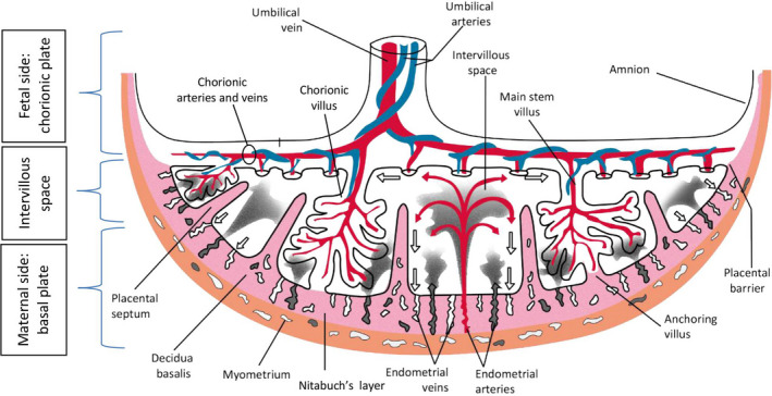
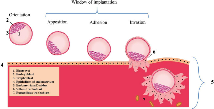
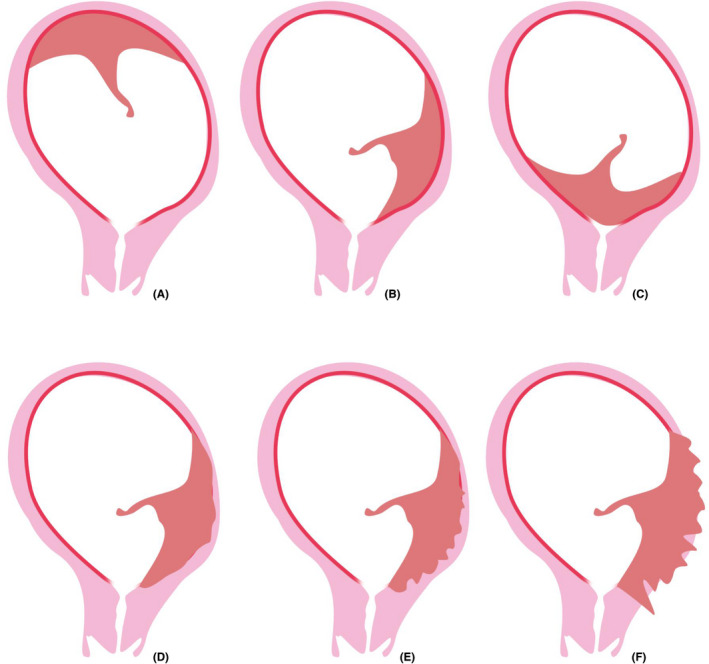
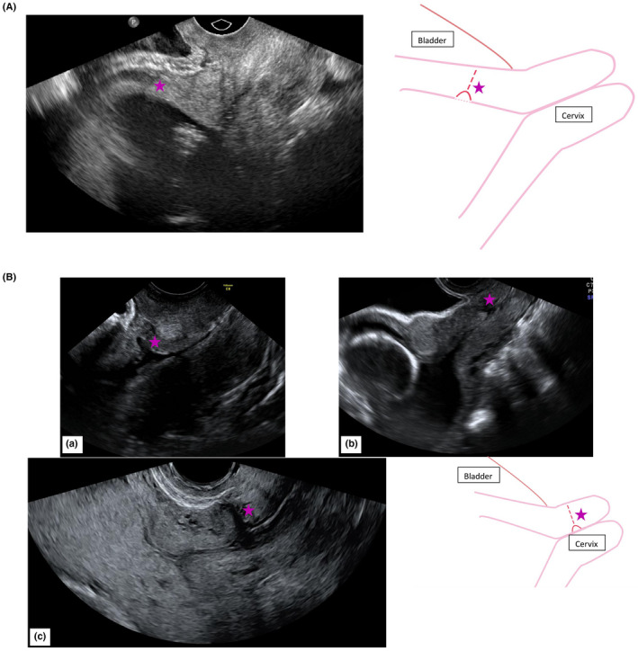
Low-lying placentas, placenta previa and abnormally invasive placentas are the most frequently occurring placental abnormalities in location and anatomy. These conditions can have serious consequences for mother and fetus mainly due to excessive blood loss before, during or after delivery. The incidence of such abnormalities is increasing, but treatment options and preventive strategies are limited. Therefore, it is crucial to understand the etiology of placental abnormalities in location and anatomy. Placental formation already starts at implantation and therefore disorders during implantation may cause these abnormalities. Understanding of the normal placental structure and development is essential to comprehend the etiology of placental abnormalities in location and anatomy, to diagnose the affected women and to guide future research for treatment and preventive strategies. We reviewed the literature on the structure and development of the normal placenta and the placental development resulting in low-lying placentas, placenta previa and abnormally invasive placentas.
Keywords: abnormally invasive placenta; low-lying placenta; placenta; placenta previa; placental abnormalities.
© 2020 The Authors. Acta Obstetricia et Gynecologica Scandinavica published by John Wiley & Sons Ltd on behalf of Nordic Federation of Societies of Obstetrics and Gynecology (NFOG).
Figures
References
-
- Rathbun KM, Hildebrand JP. Placenta abnormalities In: StatPearls. Treasure Island (FL): StatPearls Publishing; 2018. - PubMed
-
- Avagliano L, Massa V and Bulfamante GP. Histology of Human Placenta. 2016. 1–16.
-
- Silver RM. Abnormal placentation: placenta previa, vasa previa, and placenta accreta. Obstet Gynecol. 2015;126:654‐668. - PubMed
Publication types
MeSH terms
LinkOut - more resources
Full Text Sources
