. 2020 Feb 27;11(1):1085.
doi: 10.1038/s41467-020-14951-4.

A comparative genomics methodology reveals a widespread family of membrane-disrupting T6SS effectors

Affiliations

A comparative genomics methodology reveals a widespread family of membrane-disrupting T6SS effectors

Chaya M Fridman et al. Nat Commun. .

Abstract

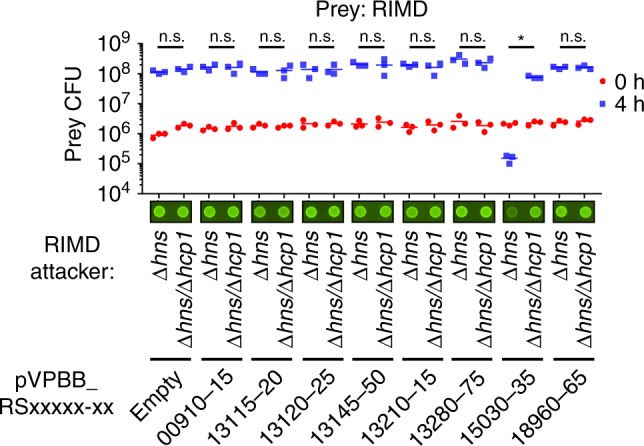
Gram-negative bacteria deliver effectors via the type VI secretion system (T6SS) to outcompete their rivals. Each bacterial strain carries a different arsenal of effectors; the identities of many remain unknown. Here, we present an approach to identify T6SS effectors encoded in bacterial genomes of interest, without prior knowledge of the effectors' domain content or genetic neighborhood. Our pipeline comprises a comparative genomics analysis followed by screening using a surrogate T6SS+ strain. Using this approach, we identify an antibacterial effector belonging to the T6SS1 of Vibrio parahaemolyticus, representing a widespread family of T6SS effectors sharing a C-terminal domain that we name Tme (Type VI membrane-disrupting effector). Tme effectors function in the periplasm where they intoxicate bacteria by disrupting membrane integrity. We believe our approach can be scaled up to identify additional T6SS effectors in various bacterial genera.


Conflict of interest statement

The authors declare no competing interests.

Figures

Fig. 1
Fig. 1. T6SS1 effector identification methodology.
a Venn diagram displaying the premise for using comparative genomics to identify T6SS1-related proteins and candidate effectors. b The first step in the T6SS1 effector identification methodology. An overview of the expected results from analyzing V. parahaemolyticus BB22OP proteins against the V. parahaemolyticus genome dataset; proteins belonging to the V. parahaemolyticus core genome are expected to have highly similar homologs in both T6SS1+ and T6SS1 genomes, whereas the closely related homologs of T6SS1 components and putative effectors should only be found in T6SS1+ genomes. c The second step in the T6SS1 effector identification methodology. Schematic representation of the V. parahaemolyticus surrogate T6SS1 platform. Candidate antibacterial T6SS1 effectors are cloned, together with a putative neighboring immunity, into an expression vector under Pbad regulation. Expression vectors are introduced into a strain with a constitutively active T6SS1 (T6SS1CA; the surrogate platform), and into a derivative mutant in which the T6SS1 is inactive (T6SS1). The ability of these attacking strains to kill a competing parental prey strain in a T6SS1-dependent manner is monitored. The parental prey strain contains the same endogenous effector/immunity (E/I) pairs as the attackers. Thus, it can antagonize their attack if they did not acquire a genuine E/I pair; however, if the expression plasmid in the attacker encodes a genuine E/I pair, then the parental prey will not be able to resist T6SS1-mediated intoxication.
Fig. 2
Fig. 2. Surrogate T6SS1 platform reveals an antibacterial effector in V. parahaemolyticus BB22OP.
Viability counts of V. parahaemolyticus RIMD 2210633 parental prey before (0 h) and after (4 h) co-incubation with surrogate platform V. parahaemolyticus RIMD 2210633 attacker strains, Δhns (T6SS1+) and Δhnshcp1 (T6SS1), on media containing L-arabinose at 30 °C (n = 3 co-cultures). Attackers harbor plasmids for the arabinose-inducible expression of the indicated V. parahaemolyticus BB22OP proteins (pVPBB_RSxxxx-xx-myc). Asterisk denotes statistical significance between samples at the 4 h timepoint by an unpaired, two-tailed Student’s t-test (P = 0.00009); n.s., no significant difference (P > 0.05). Images of representative spots of bacterial co-cultures, in which the indicated attacker strains were mixed with a parental RIMD 2210633 strain constitutively expressing GFP, are shown. Survival of GFP-expressing prey was qualitatively assessed by monitoring GFP fluorescence. Source data are provided as a source data file.
Fig. 3
Fig. 3. Tme/i1 are an antibacterial BB22OP T6SS1 E/I pair.
a Gene structure of tme/i1. Genes are represented by arrows indicating the direction of translation. White arrows denote genes unrelated to T6SS. Locus tags (VPBB_RSxxxxx) are shown above. b Expression (cells) and secretion (media) of C-terminally Flag-tagged Tme1 from T6SS1+hns) and T6SS1hnshcp1) V. parahaemolyticus BB22OP strains were detected by immunoblotting using anti-Flag antibodies. Tme1-Flag was expressed from an arabinose-inducible plasmid (pTme1). Loading control (LC), visualized as Ponceau S stained membrane, is shown for total protein lysates. The experiment was independently repeated three times with similar results. Results from a representative experiment are shown. c, d Viability counts of V. parahaemolyticus BB22OP prey before (0 h) and after (4 h) co-incubation with the indicated V. parahaemolyticus BB22OP attackers on media containing L-arabinose at 30 °C (n = 3 co-cultures). Δhns was used as the parental T6SS1+ strain, and Δhnshcp1 was used as a T6SS1 control. In (c), the prey contains an empty expression vector (pEmpty) or a vector for the arabinose-inducible expression of Tmi1 (pTmi1). In (d), attackers contain an empty expression vector (pEmpty) or a vector for the arabinose-inducible expression of Tme1 (pTme1). Asterisks denote statistical significance between samples at the 4 h timepoint by an unpaired, two-tailed Student’s t-test (*P < 0.005; **P < 0.0005; ***P < 0.00005). Source data are provided as a source data file.
Fig. 4
Fig. 4. Tme1 contains a domain that defines a widespread family of T6SS effectors.
a Conserved motif found in the C-terminal region of Tme1 (Tme) is illustrated using WebLogo 3, based on multiple sequence alignment of Tme1-homologous proteins. Cyan ovals above the WebLogo denote conserved amino acids. Secondary structure prediction (by Jpred) and transmembrane helix (TMH) prediction (by Phobius) are provided below. Alpha helices are denoted by blue cylinders, and a beta strand by an orange arrow. b Phylogenetic distribution of Tme domains constructed using the Neighbor-joining method. Tme1 and Tme2 are denoted by green and magenta circles, respectively. The scale bar represents the number of amino acid substitutions per site. c Domain architecture of Tme-containing proteins. Domain sizes are not to scale. d Pie chart of genes found immediately upstream of Tme-encoding genes. Genes encoding known T6SS structural or accessory components are indicated by their name. “Others” denotes instances where no T6SS-related genes are transcribed in the same direction as the Tme-encoding gene. The percentage of occurrences of each gene is listed next to its name.
Fig. 5
Fig. 5. Tme/i2 are an antibacterial T9109 T6SS1 E/I pair.
a Gene structure of tme/i2. Genes are represented by arrows indicating the direction of translation. White arrows denote genes unrelated to T6SS. Locus tags (PO79_RSxxxxx) are shown above. b Expression (cells) and secretion (media) of C-terminally Myc-tagged Tme2 from T6SS1+ (WT) and T6SS1hcp1) V. parahaemolyticus T9109 strains grown in the presence of phenamil (20 µM) were detected by immunoblotting using anti-Myc antibodies. Tme2-Myc was expressed from an arabinose-inducible plasmid (pTme2). Loading control (LC), visualized as trihalo compounds’ fluorescence of the immunoblot membrane, is shown for total protein lysates. The experiment was independently repeated three times with similar results. Results from a representative experiment are shown. c, d Viability counts of V. parahaemolyticus T9109 prey before (0 h) and after (4 h) co-incubation with the indicated V. parahaemolyticus T9109 attackers on media containing L-arabinose at 30 °C (n = 3 co-cultures). Δhcp1 was used as a T6SS1 control. In c, the prey contains an empty expression vector (pEmpty) or a vector for arabinose-inducible expression of Tmi2 (pTmi2). In d, attackers contain an empty expression vector (pEmpty) or a vector for the arabinose-inducible expression of Tme2 (pTme2). Asterisks denote the statistical significance between samples at the 4 h timepoint by an unpaired, two-tailed Student’s t-test (*P < 0.05; **P < 0.005; ***P < 0.001). Source data are provided as a source data file.
Fig. 6
Fig. 6. Tme effectors disrupt membrane integrity upon delivery to the periplasm.
a, b Toxicity of Tme in bacteria. Growth of E. coli BL21 (DE3) containing arabinose-inducible vectors for the expression of cytoplasmic (cyto) or periplasmic (peri) versions of Tme1 (a) and Tme2 (b). c, d Rescue of Tme-mediated toxicity by cognate Tmi. Growth of E. coli BL21 (DE3) containing an arabinose-inducible vector for the expression of periplasmic Tme1 (c) or Tme2 (d) together with an arabinose-inducible vector for the expression of Tmi1 or Tmi2. Empty expression vectors were used as the control. In ad, data represent the mean ± S.D. (n = 4). Arrows denote the time at which L-arabinose was added. e Tme effectors dissipate membrane potential. E. coli BL21 (DE3) expressing periplasmic Tme1, Tme2, or the V. cholerae pore-forming effector VasX (used as a positive control) from arabinose-inducible vectors, were analyzed using flow cytometry following staining using the BacLight Membrane Potential Kit. The red/green fluorescence ratio of the dye DiOC2(3) was calculated for each condition. CCCP was used as a positive control. Data shown in the left panel represent the mean ± S.D. of four independent experiments. Asterisks denote statistical significance compared to pEmpty samples by one-way repeated measures ANOVA with the Dunnett test (*P < 0.01; **P < 0.001; ***P < 0.0005). Data shown in the right panel represent the distribution of red/green ratios for cells analyzed in one of the experiments shown on the left. f Tme effectors increase membrane permeability. E. coli cultures, like in (e), were stained with the membrane-impermeable, intercalating DNA dye propidium iodide (PI) and analyzed using flow cytometry. Pre-treatment with ethanol (EtOH) was used as a positive control. Data shown in the left panel represent the geometric mean fluorescence intensity (MFI) ± S.D. of three repeats from a representative experiment. Asterisks denote statistical significance compared to pEmpty samples by one-way repeated measures ANOVA with the Dunnett test (**P < 0.001; ***P < 0.0005). Data shown in the right panel represent the distribution of PI fluorescence intensities for cells analyzed in one repeat of the experiment shown on the left. Source data are provided as a source data file.

References

    1. Boyer, F., Fichant, G., Berthod, J., Vandenbrouck, Y. & Attree, I. Dissecting the bacterial type VI secretion system by a genome wide in silico analysis: what can be learned from available microbial genomic resources? BMC Genomics10, (2009). - PMC - PubMed
    1. Pukatzki S, et al. Identification of a conserved bacterial protein secretion system in Vibrio cholerae using the Dictyostelium host model system. Proc. Natl Acad. Sci. USA. 2006;103:1528–1533. doi: 10.1073/pnas.0510322103. - DOI - PMC - PubMed
    1. Mougous JD, et al. A virulence locus of Pseudomonas aeruginosa encodes a protein secretion apparatus. Science. 2006;312:1526–1530. doi: 10.1126/science.1128393. - DOI - PMC - PubMed
    1. Pukatzki S, Ma AT, Revel AT, Sturtevant D, Mekalanos JJ. Type VI secretion system translocates a phage tail spike-like protein into target cells where it cross-links actin. Proc. Natl Acad. Sci. USA. 2007;104:15508–15513. doi: 10.1073/pnas.0706532104. - DOI - PMC - PubMed
    1. Ray A, et al. Type VI secretion system MIX‐effectors carry both antibacterial and anti‐eukaryotic activities. EMBO Rep. 2017;18:e201744226. doi: 10.15252/embr.201744226. - DOI - PMC - PubMed

Publication types

MeSH terms

LinkOut - more resources