. 2020 Mar 5;106(3):338-355.
doi: 10.1016/j.ajhg.2020.01.018. Epub 2020 Feb 27.

Opposite Modulation of RAC1 by Mutations in TRIO Is Associated with Distinct, Domain-Specific Neurodevelopmental Disorders

Affiliations

Opposite Modulation of RAC1 by Mutations in TRIO Is Associated with Distinct, Domain-Specific Neurodevelopmental Disorders

Sónia Barbosa et al. Am J Hum Genet. .

Abstract

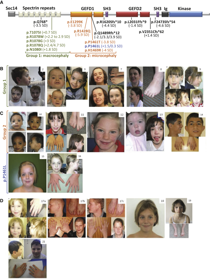
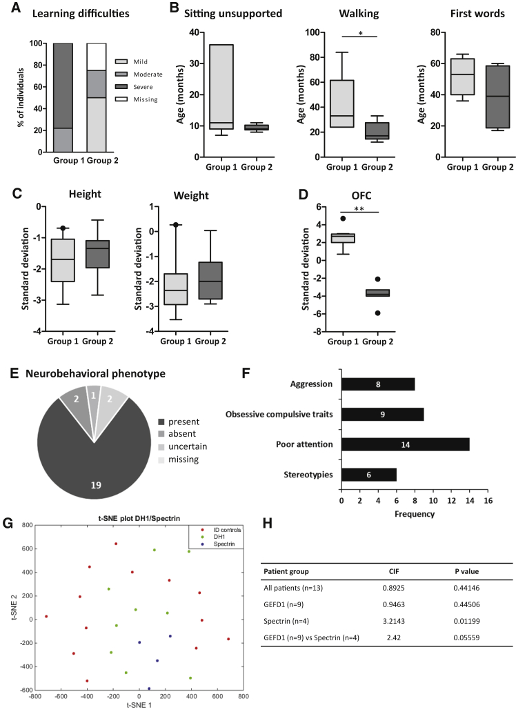
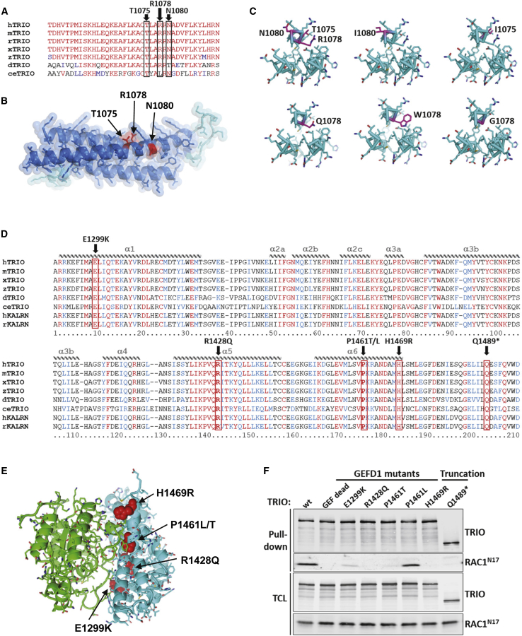
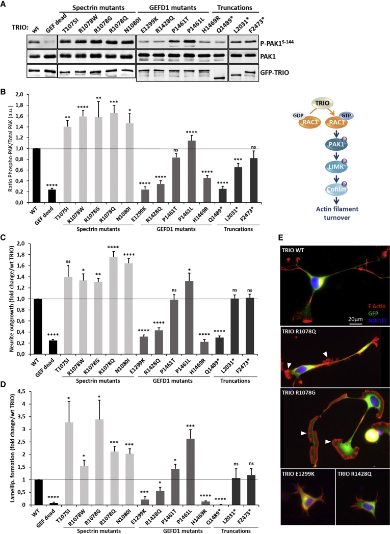
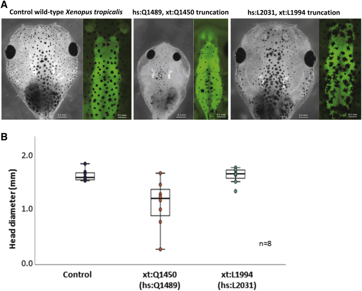
The Rho-guanine nucleotide exchange factor (RhoGEF) TRIO acts as a key regulator of neuronal migration, axonal outgrowth, axon guidance, and synaptogenesis by activating the GTPase RAC1 and modulating actin cytoskeleton remodeling. Pathogenic variants in TRIO are associated with neurodevelopmental diseases, including intellectual disability (ID) and autism spectrum disorders (ASD). Here, we report the largest international cohort of 24 individuals with confirmed pathogenic missense or nonsense variants in TRIO. The nonsense mutations are spread along the TRIO sequence, and affected individuals show variable neurodevelopmental phenotypes. In contrast, missense variants cluster into two mutational hotspots in the TRIO sequence, one in the seventh spectrin repeat and one in the RAC1-activating GEFD1. Although all individuals in this cohort present with developmental delay and a neuro-behavioral phenotype, individuals with a pathogenic variant in the seventh spectrin repeat have a more severe ID associated with macrocephaly than do most individuals with GEFD1 variants, who display milder ID and microcephaly. Functional studies show that the spectrin and GEFD1 variants cause a TRIO-mediated hyper- or hypo-activation of RAC1, respectively, and we observe a striking correlation between RAC1 activation levels and the head size of the affected individuals. In addition, truncations in TRIO GEFD1 in the vertebrate model X. tropicalis induce defects that are concordant with the human phenotype. This work demonstrates distinct clinical and molecular disorders clustering in the GEFD1 and seventh spectrin repeat domains and highlights the importance of tight control of TRIO-RAC1 signaling in neuronal development.

Keywords: autism; intellectual disability; macrocephaly; microcephaly.


Conflict of interest statement

The authors declare no competing interests.

Figures

Figure 1
Figure 1
Pathogenic TRIO Variants Found in Individuals with Neurodevelopmental Disorders (A) Schematic representation of TRIO domains with annotated missense and nonsense mutations identified in affected individuals. Patient groups 1 and 2 are highlighted in green and orange, respectively. Missense mutation p.Pro1461Leu is in blue so that it is differentiated from the other GEFD1 mutants (see text for details). Nonsense variants are written in black. The numbers in brackets correspond to the OFC of each affected individual (+/−SD). (B) Clinical photographs of individuals carrying an alteration in the seventh spectrin repeat domain of TRIO (group 1). Individuals 2 and 3 have the p.Arg1078Trp variant, individuals 7 and 8 have the p.Arg1078Gln variant, and individual 9 has the p.Asn1080Ile variant. (C) Clinical photographs of individuals carrying an alteration in the GEFD1 of TRIO (group 2 and the p.Pro1461Leu variant). Individuals 10–16 harbor the following variants: (10) p.Glu1299Lys, (11 and 12) p.Arg1428Gln, (13) p.Pro1461Thr, (14) p.His1469Arg, (15 and 16) p.Pro1461Leu. (D) Clinical photographs of individuals carrying nonsense mutations: individuals 17a–17c all carry the p.Gln1489Argfs12 variant and are related; patient 17a is the daughter of individual 17b. Individuals 17b and 17c are brothers. Individuals 18, 19, and 21 carry the nonsense variants p.Gln768, p.Arg1620Serfs10, and p.Val2351Cysfs62, respectively.
Figure 2
Figure 2
Individuals Harboring Variants in Either GEFD1 or Spectrin Domains Show Distinct Neurodevelopmental Phenotypes (A) Percentage of affected individuals with different levels of learning difficulties in either spectrin domain (group 1) or GEFD1 (group 2) mutation cases. 75% of group 2 individuals have mild-moderate learning difficulties. 78% of group 1 individuals have severe learning difficulties. (B) Early developmental milestones (sitting unsupported, walking, and first words) are delayed in both group 1 and group 2 individuals, but group 1 individuals are affected more severely. For statistical analysis, refer to Table S3. Walking: p = 0.035. (C) Height and weight standard deviations from mean in either group 1 or group 2 individuals. The median height for group 1 individuals with spectrin mutations was −1.69 SD, and for group 2 individuals with GEFD1 mutations it was −1.34 SD. 33% of the individuals in group 1 and 14% of individuals in group 2 had a height SD of greater than −2 SD, which is often considered as short stature. Weight SD was also reduced in both groups. (D) Microcephaly was seen in 100% of group 2 individuals, who had a mean OFC of −3.82 SD (median OFC of −3.8 SD). Individuals within group 1 present with macrocephaly. 78% had an OFC greater than 2 SD from the mean; mean OFC was +2.6 SD, median OFC was + 2.7 SD, and OFC range was between +0.7 SD and +4.7 SD. ∗∗p < 0.001 (Table S3) (E) A neurobehavioral phenotype was observed in 19 out of the 24 (79%) individuals described in this study. (F) Recurrent behavioral features include stereotypies (6/24 patients), poor attention (14/24 individuals), obsessive-compulsive traits (9/24 individuals), and aggression (8/24 individuals). In total, 2/24 were identified as social or friendly. (G and H) Quantitative facial phenotyping. t-distributed stochastic neighbor embedding (t-SNE) plot of the vectors of individuals with alterations in GEFD1 and the seventh spectrin domain and the matched controls with intellectual disability. The statistically significant clustering of individuals with mutations in the spectrin domain indicates a recognizable facial phenotype.
Figure 3
Figure 3
Mapping of the Mutation Sites on the 3D Structure of the TRIO Spectrin 7 Repeat Domain and GEFD1 (A) Species conservation of the residues in TRIO’s seventh spectrin repeat, which is mutated in neurodevelopmental diseases. Identical residues are labeled in red, and similar residues are in blue. The positions of the residues p.Thr1075, p.Arg1078, and p.Asn1080 are boxed in black and indicated on top of the sequence, which encompasses amino acids 1053 to 1091, corresponding to the second α helix of the spectrin repeat. Represented species are Homo sapiens (h), Mus musculus (m), Rattus norvegicus (r), Xenopus laevis (x), Danio rerio (z), Drosophila melanogaster (d), and Caenorhabditis elegans (ce). (B) Lateral view of the structural model of the seventh spectrin repeat of TRIO. The spectrin domain was modeled based on the crystal structures of human beta2-spectrin (PDB ID 3EDV30) with our sequence alignment and SWISS-MODEL server. Parts of the structure that were modeled with high and low confidence are in blue and cyan, respectively. Mutations within the spectrin domain seen in affected individuals are indicated in red. (C) Axial view of the cross section of the structure at the site of the mutations. Mutations are indicated in magenta. (D) Sequence alignment of the RAC1-specific DH1 domain of TRIO (and KALIRIN) across evolution. Identical residues are labeled in red and similar residues are labeled in blue. The α helices are depicted schematically on top of the sequence alignment. The positions of the alterations p.Glu1299Lys, p.Arg1428Gln, p.Pro1461Leu, p.Pro1461Thr, and p.His1469Arg are indicated in bold and boxed in red. Each mutation affects a highly conserved residue within helices α-1, α-5, and α-6, which make contact with the target GTPase RAC1. Represented species are Homo sapiens (h), Mus musculus (m), Rattus norvegicus (r), Xenopus laevis (x), Danio rerio (z), Drosophila melanogaster (d), and Caenorhabditis elegans (ce). (E) Structure of the DH1 domain of TRIO (cyan) in complex with the small GTPase substrate RAC1 (green). Mutations within the DH1 domain seen in affected individuals are indicated in red and can be seen to occur at the protein-substrate interface. The complex was modeled with the crystal structures of DH1 (PDB: 1NTY) and the complex with substrate (PDB: 1KZ7). Figures of the protein structures were generated with PyMol. (F) Most of the GEFD1 mutants are affected in their ability to bind to RAC1N17 (DN). Immunoblot analysis of a Streptavidin pulldown assay of biotinylated TRIO variants. HEK293T cells were transfected with the indicated biotinylated GFP-TRIO variants and RAC1N17. TRIO was pulled down with Streptavidin beads, and the co-precipitating RAC1N17 was detected with a RAC1 antibody.
Figure 4
Figure 4
The Spectrin Mutants of TRIO Enhance RAC1 Signaling, Neurite Outgrowth, and Lamellipodia Formation in N1E-115 Cells, Whereas the GEFD1 Mutants Are Mostly Impaired in These Processes (A) Immunoblot analysis of HEK293T cell lysates transfected with the indicated GFP-TRIO variants and detected with an anti-GFP antibody (lower panel). PAK1 phosphorylation amounts are detected with a phospho-Ser144 PAK1 antibody (upper panel) and compared to total PAK1 amounts detected with a PAK1 antibody (middle panel). (B) Quantification of the ratio of phospho-PAK1 amounts over total PAK1 expression. PAK1 phosphorylation is used as a readout for the activation of the RAC1 signaling cascade. Data are presented as the mean ± SEM of at least five independent experiments. (C) Quantification of the neurite outgrowth induced by WT or mutant TRIO. Neurite outgrowth is monitored on the basis of the number of cells harboring an extension of at least twice the length of the soma. Data are presented as n-fold change over WT TRIO, which was arbitrarily set to 1. Data are presented as the mean ± SEM of at least five independent experiments. (D) Quantification of lamellipodia formation induced by WT or mutant TRIO. Data are presented as n-fold change over WT TRIO, which was arbitrarily set to 1. Data are presented as the mean ± SEM of at least five independent experiments. Statistical analysis in (B), (C), and (D) were made by one-way ANOVA followed by Dunnett’s test. Asterisks indicate datasets significantly different from WT (p < 0.05, ∗∗p < 0.01, ∗∗∗p < 0.001, and ∗∗∗∗p < 0.0001). (E) Micrographs of N1E-115 cells transfected with the indicated GFP-TRIO variants (green); rhodamine-phalloidin and Hoechst stained the actin (red) and nuclei (blue), respectively. Representative images for each variant type are presented. White arrowheads point to lamellipodia. Scale bar: 20 μm.
Figure 5
Figure 5
Truncation in the TRIO GEFD1 Reduces Head Size in X. tropicalis (A) X. tropicalis eggs from a single female for each of two biological replicates (with parents from different genetic backgrounds) were fertilized with frozen sperm and injected at the one-cell stage with CRISPR-Cas targeting the genome as shown. Representative micrographs for each condition are shown (black and white photos) (all tadpoles are shown in Figure S3). Performing the GEFD1 and GEFD2 frameshift in a tubb2B.GFP X. tropicalis showed that forebrain deformities had occurred in 6/8 GEFD1 truncation tadpoles, which was not the case for GEFD2 frameshift. Representative micrographs for each condition are shown (color photos) (all tadpoles are shown in Figure S4). (B) Comparing the head diameter of the tadpoles carrying truncations in the GEFD1 (hs: Gln 1489 = xt Gln 1450) and GEFD2 (hs: Leu 2031 = xt Leu 1994) domains showed that GEFD1 truncations caused microcephaly, whereas GEFD2 truncations had no effect. Quantification is shown for eight individuals measured (all tadpoles are shown in Figure S3).

References

    1. Govek E.E., Newey S.E., Van Aelst L. The role of the Rho GTPases in neuronal development. Genes Dev. 2005;19:1–49. - PubMed
    1. Tolias K.F., Duman J.G., Um K. Control of synapse development and plasticity by Rho GTPase regulatory proteins. Prog. Neurobiol. 2011;94:133–148. - PMC - PubMed
    1. Debant A., Serra-Pagès C., Seipel K., O’Brien S., Tang M., Park S.H., Streuli M. The multidomain protein Trio binds the LAR transmembrane tyrosine phosphatase, contains a protein kinase domain, and has separate rac-specific and rho-specific guanine nucleotide exchange factor domains. Proc. Natl. Acad. Sci. USA. 1996;93:5466–5471. - PMC - PubMed
    1. Blangy A., Vignal E., Schmidt S., Debant A., Gauthier-Rouvière C., Fort P. TrioGEF1 controls Rac- and Cdc42-dependent cell structures through the direct activation of rhoG. J. Cell Sci. 2000;113:729–739. - PubMed
    1. Bellanger J.M., Lazaro J.B., Diriong S., Fernandez A., Lamb N., Debant A. The two guanine nucleotide exchange factor domains of Trio link the Rac1 and the RhoA pathways in vivo. Oncogene. 1998;16:147–152. - PubMed

Publication types

Substances