

Clinical and computed tomographic imaging features of novel coronavirus pneumonia caused by SARS-CoV-2
- PMID: 32109443
- PMCID: PMC7102535
- DOI: 10.1016/j.jinf.2020.02.017
Clinical and computed tomographic imaging features of novel coronavirus pneumonia caused by SARS-CoV-2
Abstract
Purpose: To investigate the clinical and imaging characteristics of computed tomography (CT) in novel coronavirus pneumonia (NCP) caused by SARS-CoV-2.
Materials and methods: A retrospective analysis was performed on the imaging findings of patients confirmed with COVID-19 pneumonia who had chest CT scanning and treatment after disease onset. The clinical and imaging data were analyzed.
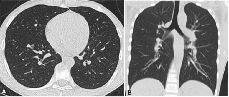
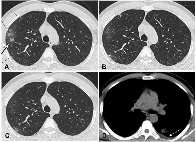
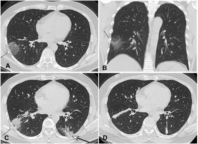
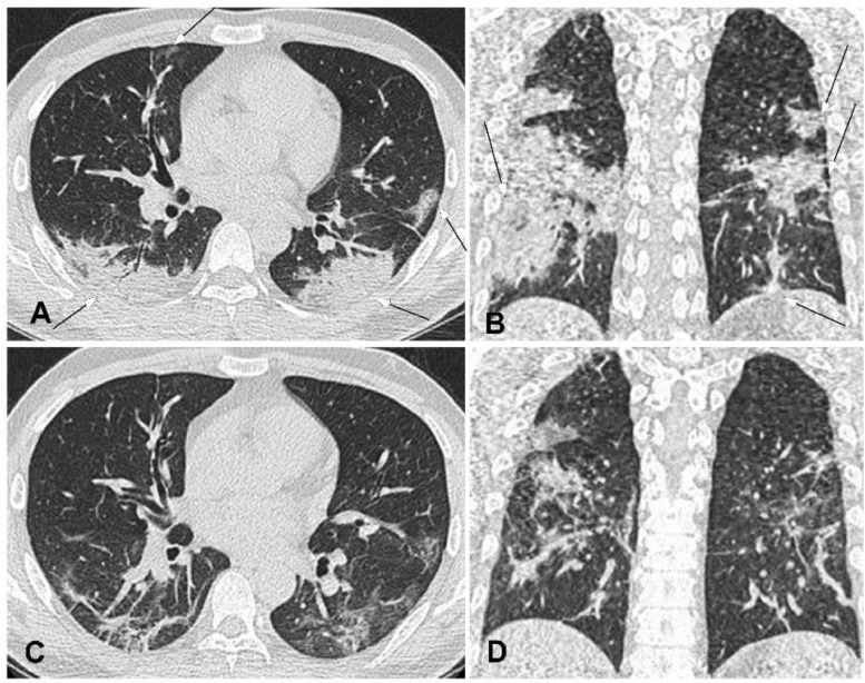
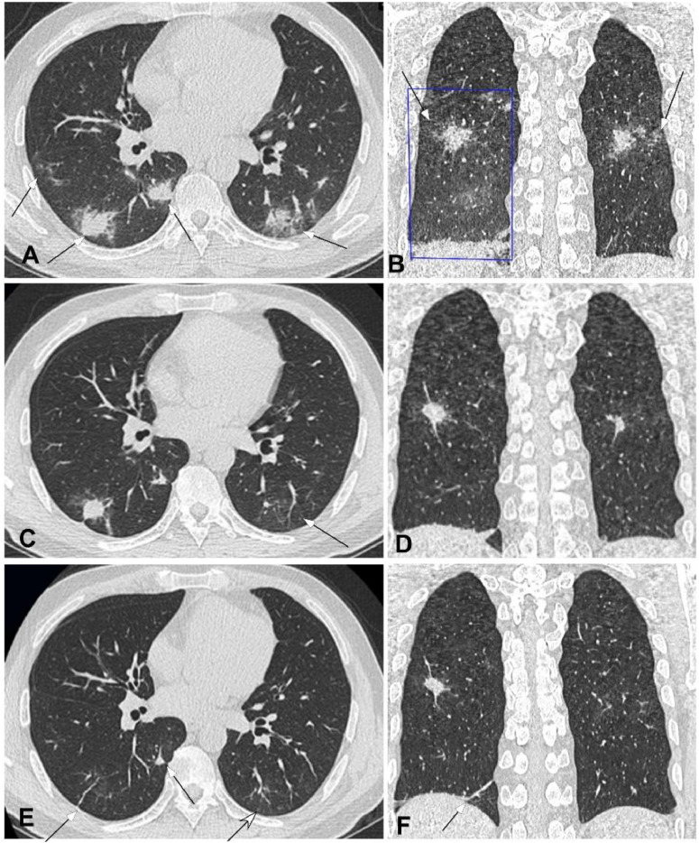
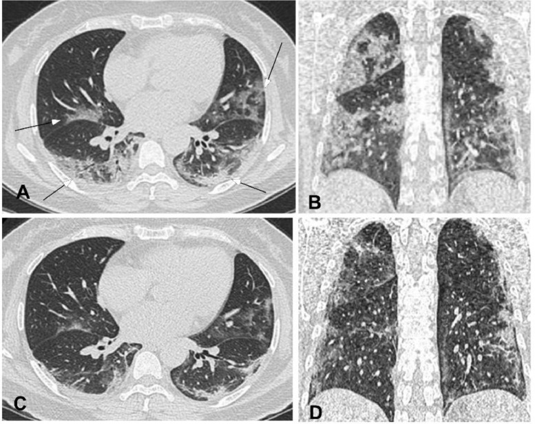
Results: Fifty patients were enrolled, including mild type in nine, common in 28, severe in 10 and critically severe in the rest three. Mild patients (29 years) were significantly (P<0.03) younger than either common (44.5 years) or severe (54.7) and critically severe (65.7 years) patients, and common patients were also significantly (P<0.03) younger than severe and critically severe patients. Mild patients had low to moderate fever (<39.1 °C), 49 (98%) patients had normal or slightly reduced leukocyte count, 14 (28%) had decreased counts of lymphocytes, and 26 (52%) patients had increased C-reactive protein. Nine mild patients were negative in CT imaging. For all the other types of NCP, the lesion was in the right upper lobe in 30 cases, right middle lobe in 22, right lower lobe in 39, left upper lobe in 33 and left lower lobe in 36. The lesion was primarily located in the peripheral area under the pleura with possible extension towards the pulmonary hilum. Symmetrical lesions were seen in 26 cases and asymmetrical in 15. The density of lesion was mostly uneven with ground glass opacity as the primary presentation accompanied by partial consolidation and fibrosis.
Conclusion: CT imaging presentations of NCP are mostly patchy ground glass opacities in the peripheral areas under the pleura with partial consolidation which will be absorbed with formation of fibrotic stripes if improved. CT scanning provides important bases for early diagnosis and treatment of NCP.
Keywords: Computed tomography; Covid-19; Imaging finding; Novel coronavirus pneumonia; SARS-CoV-2.
Copyright © 2020 The British Infection Association. Published by Elsevier Ltd. All rights reserved.
Conflict of interest statement
Declaration of Competing Interest None.
Figures
Comment in
-
The evolution of CT characteristics in the patients with COVID-19 pneumonia.J Infect. 2020 Jun;80(6):e29. doi: 10.1016/j.jinf.2020.03.014. Epub 2020 Mar 19. J Infect. 2020. PMID: 32201155 Free PMC article. No abstract available.
-
Observation and analysis of 26 cases of asymptomatic SARS-COV2 infection.J Infect. 2020 Jul;81(1):e69-e70. doi: 10.1016/j.jinf.2020.03.028. Epub 2020 Apr 3. J Infect. 2020. PMID: 32251687 Free PMC article. No abstract available.
-
TEMPORARY REMOVAL: Characteristics of deaths amongst health workers in China during the outbreak of COVID-19 infection.J Infect. 2020 Jul;81(1):147-178. doi: 10.1016/j.jinf.2020.03.030. Epub 2020 Apr 8. J Infect. 2020. PMID: 32277966 Free PMC article. No abstract available.
-
High-resolution computed tomographic imaging disclosing COVID-19 pneumonia: A powerful tool in diagnosis.J Infect. 2020 Aug;81(2):318-356. doi: 10.1016/j.jinf.2020.03.047. Epub 2020 Apr 10. J Infect. 2020. PMID: 32283148 Free PMC article. No abstract available.
References
MeSH terms
LinkOut - more resources
Full Text Sources
Other Literature Sources
Research Materials
Miscellaneous
