. 2020 Jun:70:109588.
doi: 10.1016/j.cellsig.2020.109588. Epub 2020 Feb 26.

Chronic WNT/β-catenin signaling induces cellular senescence in lung epithelial cells

Affiliations

Chronic WNT/β-catenin signaling induces cellular senescence in lung epithelial cells

Mareike Lehmann et al. Cell Signal. 2020 Jun.

Abstract

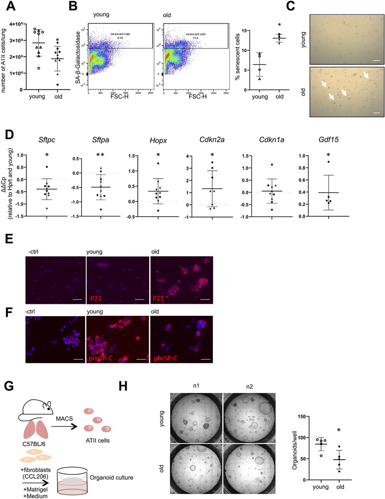
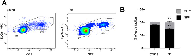
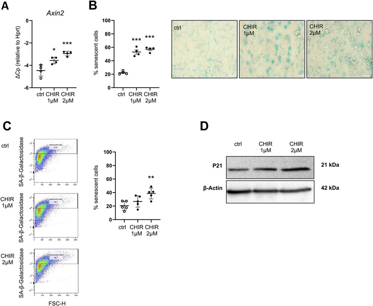
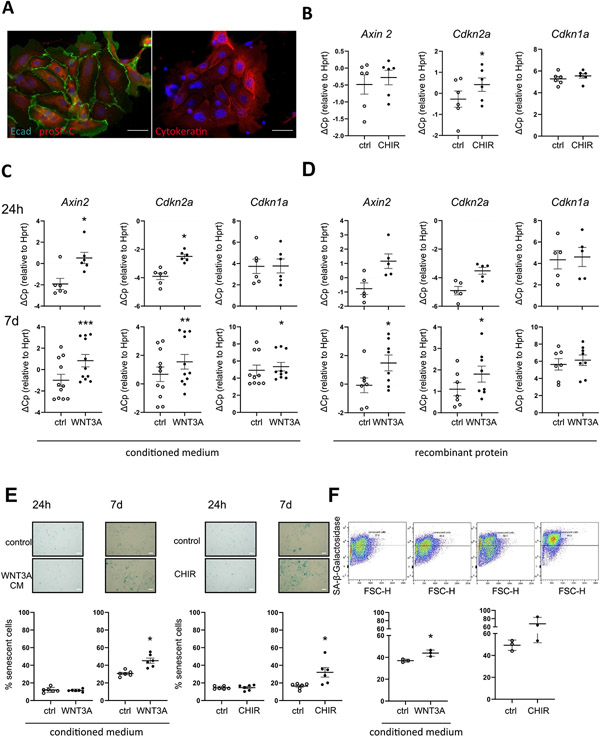
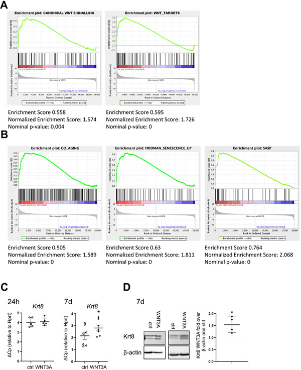
The rapid expansion of the elderly population has led to the recent epidemic of age-related diseases, including increased incidence and mortality of chronic lung diseases, such as Idiopathic Pulmonary Fibrosis (IPF). Cellular senescence is a major hallmark of aging and has a higher occurrence in IPF. The lung epithelium represents a major site of tissue injury, cellular senescence and aberrant activity of developmental pathways such as the WNT/β-catenin pathway in IPF. The potential impact of WNT/β-catenin signaling on alveolar epithelial senescence in general as well as in IPF, however, remains elusive. Here, we characterized alveolar epithelial cells of aged mice and assessed the contribution of chronic WNT/β-catenin signaling on alveolar epithelial type (AT) II cell senescence. Whole lungs from old (16-24 months) versus young (3 months) mice had relatively less epithelial (EpCAM+) but more inflammatory (CD45+) cells, as assessed by flow cytometry. Compared to young ATII cells, old ATII cells showed decreased expression of the ATII cell marker Surfactant Protein C along with increased expression of the ATI cell marker Hopx, accompanied by increased WNT/β-catenin activity. Notably, when placed in an organoid assay, old ATII cells exhibited decreased progenitor cell potential. Chronic canonical WNT/β-catenin activation for up to 7 days in primary ATII cells as well as alveolar epithelial cell lines induced a robust cellular senescence, whereas the non-canonical ligand WNT5A was not able to induce cellular senescence. Moreover, chronic WNT3A treatment of precision-cut lung slices (PCLS) further confirmed ATII cell senescence. Simultaneously, chronic but not acute WNT/β-catenin activation induced a profibrotic state with increased expression of the impaired ATII cell marker Keratin 8. These results suggest that chronic WNT/β-catenin activity in the IPF lung contributes to increased ATII cell senescence and reprogramming. In the fibrotic environment, WNT/β-catenin signaling thus might lead to further progenitor cell dysfunction and impaired lung repair.

Keywords: ATII cells; Aging; Cellular senescence; IPF; WNT signaling.


Figures

Fig. 1.
Fig. 1.
Phenotype of ATII cells in aged mice. (A-H) Lungs from young (6–21 weeks) or old (16–24 months) mice were harvested and ATII cells were isolated. (A) Total number of isolated ATII cells per mouse, n = 10. (B) Freshly isolated ATII cells were analyzed for SA-ß-galactosidase activity by FACS n = 4 or (C) by conventional SA-ß-galactosidase staining 2 days after plating, image representative of n = 4, arrows depict positive cells, Size bar represents 50 μm. (D) Freshly isolated ATII cells were analyzed for alveolar epithelial cell and senescence markers by qPCR. Values were normalized to Hprt and corresponding young controls. n = 5–10. (E-F) Cytospins of freshly isolated ATII cells were stained for (E) P21 or (F) proSP-C protein. Image representative of a n = 3, Size bar represents 50 μm. (G-H) Freshly isolated ATII cells were combined with Matrigel and CCL206 fibroblasts and used for an organoid assay as outlined in (G) and representative pictures are shown at day 14 of organoid differentiation in (H) as well as quantification of numbers of organoids per well, n = 5. Data are presented as mean ± s.d. Circles represent C57BL6/J mice, triangles represent C57BL6/N mice. Significance was assessed with one sample t-test compared to a hypothetical value of 0 (D) or Mann Whitney-test (A, B, H) Significance: *P < .05, **P .01.
Fig. 2.
Fig. 2.
WNT activity is increased in ATII cells from aged mice. (A-B) Lungs from young (3 months) or old (18 months) WNT GFP mice were harvested and ATII cells were isolated and analyzed for WNT activity (GFP+) by FACS. Data are presented as mean ± s.d. n = 6 old, n = 14 young. Significance was assessed with a two-way Anova followed by Sidak's multiple comparison test. Significance: **P < .01.
Fig. 3.
Fig. 3.. Chronic WNT stimulation induces cellular senescence in MLE12 cells.
(A-D) MLE12 lung epithelial cells were treated with 1 μM CHIR or 2 μM CHIR for 7d (A) qPCR analysis for WNT target gene Axin2 normalized to Hprt levels was perfomed. n = 4. (B) SA-β-galactosidase activity was measured by conventional staining after 7d, representative of n = 4. Size bar represents 50 μm. (C) SA-β-galactosidase activity was measured by FACS-based staining after 7d, n = 5. (D) Western Blot for P21 was performed, β-actin was used as a loading control. Blot is representative of n = 4. Data are presented as mean ± s.d. Significance was assessed with a one-way Anova followed by Tukey's multiple comparison test. Significance: *P < .05, **P < .01, ***P < .001.
Fig. 4.
Fig. 4.. Chronic WNT stimulation induces cellular senescence in pmATII cells.
(A) ATII cells were stained by Immunofluorescence for pro Surfactant Protein-C (proSP-C), E-Cadherin (Ecad) or Cytokeratin. (B) ATII cells were treated with 1 μM CHIR for 7d, n = 6, (C) conditioned medium from WNT3A-overexpressing L-cells (WNT3A CM; 1:1) for 24 h (n = 6) or 7 days (n = 10) (D) recombinant mWNT3A for 24 h (n = 5) and 7d (n = 8). (B-D) qPCR analysis for WNT target gene Axin2 and senescence markers Cdkn1a and Cdkn2a normalized to Hprt levels was performed. (E-F) ATII cells were treated with 1 μM CHIR or WNT3A CM and SA-β-galactosidase activity was measured by (E) conventional staining after 24 h and 7d, n = 6 or by (F) FACS-based staining after 7d, n = 3. Significance was assessed by Wilcoxon matched-pair signed rank test (B-D) and Student's t-test (E-F). Significance: *p < .05, **P < .01.
Fig. 5.
Fig. 5.. Chronic WNT stimulation induces and cellular senescence in epithelial cells.
(A-C)Preciscion cut lung slices (PCLS) were prepared from young mice and treated with (A) 2 μM CHIR for 7 days. n = 5, or (B) conditioned medium from WNT3A-overexpressing L-cells (WNT3A CM; 1:1) for 7 days, n = 3. (A-B) qPCR analysis for WNT target gene Axin2 and senescence markers Cdkn2a and Cdkn1a was performed and normalized to Hprt levels. (C) Representative images of immunofluorescence staining for P21 and CDH1 (E-CAD) in PCLS prepared from young mice and treated with WNT3A CM for 7 days. Fluorescent images represent a 400 × magnification. The scale bar represents 50 μm. Representative of n = 3. Significance was assessed by paired Student's t-test (A-B). Significance: *p < .05, **P < .01.
Fig. 6.
Fig. 6.. Chronic WNT stimulation induces Keratin 8 (Krt8) and cellular senescence in epithelial cells.
(A-B) Gene set enrichment analysis was performed on a published RNA sequencing dataset from hATII cells from Donor/IPF patients GSE94555 (Xu et al., 2016, JCI Insight). The dataset was tested for the enrichment of (A) WNT/β-catenin (GO: 0060070 and https://web.Stanford.edu/group/nusselab/cgi-bin/wnt/target_-genes) or (B) for aging (GO:0007568), senescence (Fridman et al., Oncogene, 2008 [40]) or senescence associated secretory phenotype (SASP; Coppé, Annu Rev. Pathol) [9]) lists. (C-D) pmATII cells were treated with WNT3A CM for 24 h (n = 5) or 7days (n = 7). (D) qPCR analysis of the fibrotic epithelial marker Krt8 normalized to Hprt(D) Two representative western blots of Krt8. Quantification of Krt8 signal over β-actin normalized to Ctrl CM is shown on the right. n = 4. Data are presented as mean ± s.d. Significance was assessed by Wilcoxon matched-pair signed rank test (C) and one sample t-test compared to a hypothetical value of 1 (D). Significance: *p < .05, **P < .01.

References

    1. Bowdish DME, The aging lung: is lung health good health for older adults? Chest 155 (2) (2019) 391–400. - PubMed
    1. Meiners S, Eickelberg O, Konigshoff M, Hallmarks of the ageing lung, Eur. Respir. J 45 (3) (2015) 807–827. - PubMed
    1. Barkauskas CE, Cronce MJ, Rackley CR, Bowie EJ, Keene DR, Stripp BR, Randell SH, Noble PW, Hogan BL, Type 2 alveolar cells are stem cells in adult lung, J. Clin. Invest 123 (7) (2013) 3025–3036. - PMC - PubMed
    1. Selman M, Pardo A, Role of epithelial cells in idiopathic pulmonary fibrosis: from innocent targets to serial killers, Proc. Am. Thorac. Soc 3 (4) (2006) 364–372. - PubMed
    1. Lederer DJ, Martinez FJ, Idiopathic pulmonary fibrosis, N. Engl. J. Med 379 (8) (2018) 797–798. - PubMed

Publication types