. 2020 Feb 19:15:1215-1228.
doi: 10.2147/IJN.S234850. eCollection 2020.

PEGylated PLGA Nanoparticle Delivery of Eggmanone for T Cell Modulation: Applications in Rheumatic Autoimmunity

Affiliations

PEGylated PLGA Nanoparticle Delivery of Eggmanone for T Cell Modulation: Applications in Rheumatic Autoimmunity

Christopher P Haycook et al. Int J Nanomedicine. .

Abstract

Background: Helper T cell activity is dysregulated in a number of diseases including those associated with rheumatic autoimmunity. Treatment options are limited and usually consist of systemic immune suppression, resulting in undesirable consequences from compromised immunity. Hedgehog (Hh) signaling has been implicated in the activation of T cells and the formation of the immune synapse, but remains understudied in the context of autoimmunity. Modulation of Hh signaling has the potential to enable controlled immunosuppression but a potential therapy has not yet been developed to leverage this opportunity.

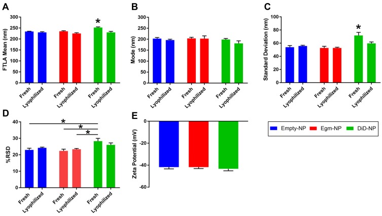
Methods: In this work, we developed biodegradable nanoparticles to enable targeted delivery of eggmanone (Egm), a specific Hh inhibitor, to CD4+ T cell subsets. We utilized two FDA-approved polymers, poly(lactic-co-glycolic acid) and polyethylene glycol, to generate hydrolytically degradable nanoparticles. Furthermore, we employed maleimide-thiol mediated conjugation chemistry to decorate nanoparticles with anti-CD4 F(ab') antibody fragments to enable targeted delivery of Egm.

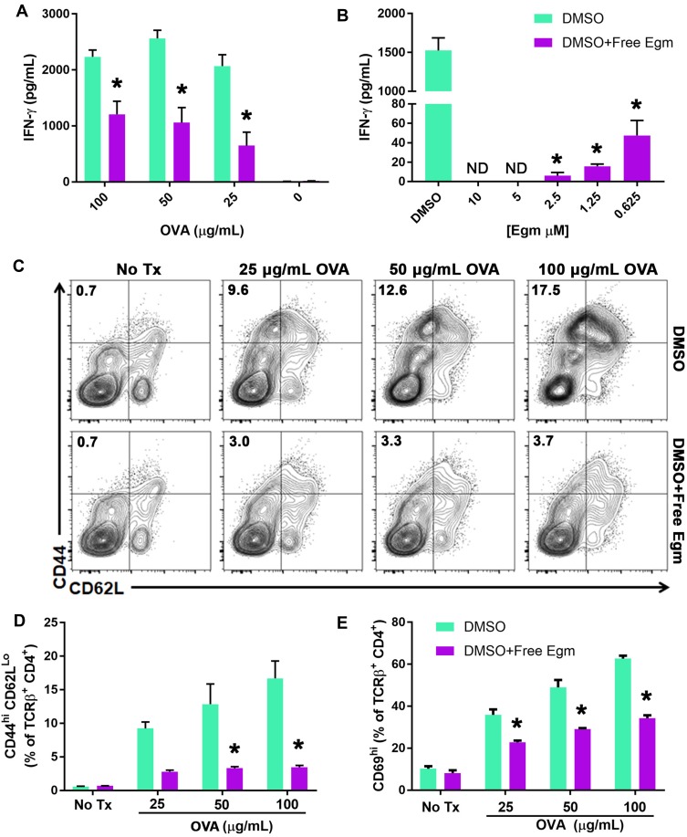
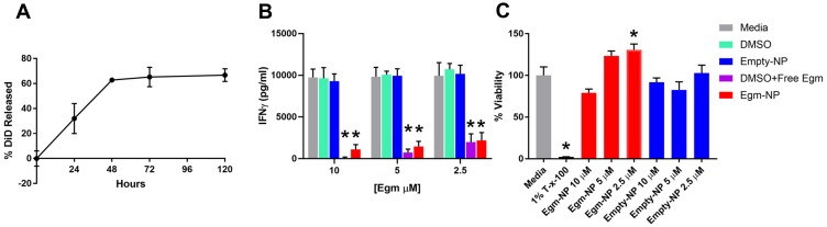
Results: Our novel delivery system achieved a highly specific association with the majority of CD4+ T cells present among a complex cell population. Additionally, we have demonstrated antigen-specific inhibition of CD4+ T cell responses mediated by nanoparticle-formulated Egm.

Conclusion: This work is the first characterization of Egm's immunomodulatory potential. Importantly, this study also suggests the potential benefit of a biodegradable delivery vehicle that is rationally designed for preferential interaction with a specific immune cell subtype for targeted modulation of Hh signaling.

Keywords: advanced delivery systems; autoimmunity; controlled release; eggmanone.


Conflict of interest statement

Professor Charles C Hong and Dr Charles H Williams report a US Patent #: US 10,329,304 B2 issued. Professor Todd D Giorgio reports patent materials and methods for the treatment of immune cells pending. The authors report no other conflicts of interest in this work.

Figures

Figure 1
Figure 1
Egm inhibits CD4+ T cell activation and cytokine responses. Notes: (A) OT-II whole splenocytes were incubated with whole OVA protein at various concentrations for 72 hrs in the presence of DMSO (vehicle) or 10 µM Egm in DMSO. (B) Egm dose response of OT-II whole splenocytes incubated with OVA323-339 peptide (50 µg/mL) for 72 hrs. IFN-γ concentration was measured via ELISA. (C) Representative flow cytometric analysis of OT-II whole splenocyte cultures incubated with OVA323-339 peptide in the presence of DMSO or 10 µM Egm in DMSO for 72 hrs. T cell activation was evaluated by analysis of CD44hiCD62Llo T cell populations (plots gated from TCRβ+CD4+ T cells). (D) Quantification of CD44hiCD62Llo T cell populations from 3 biological replicates. (E) Quantification of CD69 expression in the presence of Egm or DMSO from 3 biological replicates. The significance of the data was evaluated via ordinary One-way ANOVA with multiple comparison test. (*P<0.05). Abbreviations: OVA, ovalbumin; DMSO, dimethyl sulfoxide; No Tx, no treatment.
Figure 2
Figure 2
PEGylated PLGA nanoparticles retain physicochemical characteristics following Egm loading. Notes: (A) FTLA mean hydrodynamic diameter of nanoparticles. (B) Mode of nanoparticle diameter distributions. (C) Standard deviation of nanoparticle diameters. (D) Calculated percent relative standard deviation of nanoparticle distributions (E) Zeta potential measures of lyophilized formulations. All measurements were performed in deionized water. All data shown represent the mean ± SEM of at least three independent batches for each formulation. The significance of the data was evaluated via ordinary One-way or Two-way ANOVA with multiple comparison test. (*P<0.05). Abbreviations: FTLA, finite-track length adjusted; empty-NP, empty nanoparticle; Egm-NP, Eggmanone-loaded nanoparticle; DiD-NP, DiD-loaded nanoparticle; SEM, standard error of the mean.
Figure 3
Figure 3
Emulsion mediated fabrication localizes Egm in nanoparticle cores. Notes: (A) Transmission electron micrographs of empty and Egm-loaded nanoparticles. No negative staining was utilized. (B) Elemental analysis and quantification of sulfur weight % and atoms % within empty and Egm-loaded nanoparticles. Inset corresponds to the energy region within the green box. (C) Nanoscale X-ray element mappings of empty nanoparticle. (D) Nanoscale X-ray element mappings of Egm-loaded nanoparticle. All images representative of corresponding nanoparticle populations. All scale bars represent 200 nm. Abbreviations: Empty-NP, empty nanoparticle; Egm-NP, Egm-loaded nanoparticle; HAADF, high-angle angular darkfield image; C, carbon; S, sulfur; O, oxygen; N, nitrogen, Cu, copper; Si, silicon; Cl, chlorine; kα, K-alpha emission line.
Figure 4
Figure 4
Nanoparticle-formulated Egm inhibits CD4+ T cell cytokine responses. Notes: (A) Normalized release profile of DiD from nanoparticles incubated in 37°C 1× PBS. (B) Interferon gamma (IFN-γ) production measured by ELISA from OT-II splenocytes stimulated with 50 µg/mL whole ovalbumin and incubated with vehicle controls, Egm in DMSO, and Egm-loaded nanoparticles for 72 hrs (n=3 biological replicates). (C) Average viability normalized to media controls of whole FVB splenocytes incubated for 72 hrs with Egm- and empty-NPs (n=3 biological replicates). No acute toxicity was observed for either particle formulation. Final Egm concentration of particle suspensions is listed. Empty particle concentrations correspond to matched polymer doses. The significance of the data was evaluated via ordinary One-way ANOVA with multiple comparison test (*p<0.05). Abbreviations: DiD, 1,1ʹ-dioctadecyl-3,3,3ʹ,3ʹ-tetramethylindodicarbocyanine, 4-chlorobenzenesulfonate salt; T-x-100, triton-x-100; empty-NP, empty nanoparticle; Egm-NP, Egm-loaded nanoparticle; DMSO, dimethyl sulfoxide.
Figure 5
Figure 5
F(ab’) fragment conjugated PEGylated PLGA nanoparticles target CD4+ T cells. Notes: (A) Flow cytometry gating strategy used to analyze the targeting specificity of anti-CD4 decorated DiD-NPs and representative staining results from 1 of 3 experiments. Full flow cytometry gating strategy for targeting experiments shown in Supplemental Figure 3. (B) Quantification of DiD positive splenocytes incubated with either undecorated, isotype control decorated, or anti-CD4 decorated fluorescent nanoparticles suspended in 1× PBS. The flow cytometry probe for CD4 staining utilized an antibody clone that recognized a different epitope of CD4 than those used to decorate nanoparticles. (C) Analysis of CD4+ T cell targeting specificity after incubation with increasing concentrations of anti-CD4-decorated DiD-NPs. Nanoparticle concentrations were determined by nanoparticle tracking analysis. The percentage of DiD+CD4+ T cells and DiD+CD8+ T cells is indicated in the upper right-hand quadrants of the flow cytometry plots. Splenocytes were derived from female C57BL/6J mice. The significance of the data was evaluated via ordinary Two-way ANOVA with multiple comparison test (*p < 0.05). Abbreviations: SSC, side scatter; FSC, forward scatter; undecorated-NP, undecorated DiD-NP; isotype-NP, isotype control antibody fragment decorated DiD-NP; α-CD4-NP, anti-CD4 antibody fragment decorated DiD-NP.

References

    1. Marder W, Vinet É, Somers EC. Rheumatic autoimmune diseases in women and midlife health. Women’s Midlife Heal. 2015;1:1–8. - PMC - PubMed
    1. Vaughan S, Dawe HR. Common themes in centriole and centrosome movements. Trends Cell Biol. 2011;21:57–66. doi:10.1016/j.tcb.2010.09.004 - DOI - PubMed
    1. De La Roche M, Ritter AT, Angus KL, et al. Hedgehog signaling controls T cell killing at the immunological synapse. Science. 2013;342:1247–1250. doi:10.1126/science.1244689 - DOI - PMC - PubMed
    1. Smelkinson M. The hedgehog signaling pathway emerges as a pathogenic target. J Dev Biol. 2017;5:14. doi:10.3390/jdb5040014 - DOI - PMC - PubMed
    1. Crompton T, Outram SV, Hager-Theodorides AL. Sonic hedgehog signalling in T-cell development and activation. Nat Rev Immunol. 2007;7:726–735. doi:10.1038/nri2151 - DOI - PubMed

MeSH terms