. 2020 Feb 4;18(1):1559325820904547.
doi: 10.1177/1559325820904547. eCollection 2020 Jan-Mar.

Alteration of Metal Elements in Radiation Injury: Radiation-Induced Copper Accumulation Aggravates Intestinal Damage

Affiliations

Alteration of Metal Elements in Radiation Injury: Radiation-Induced Copper Accumulation Aggravates Intestinal Damage

Li Zhong et al. Dose Response. .

Abstract

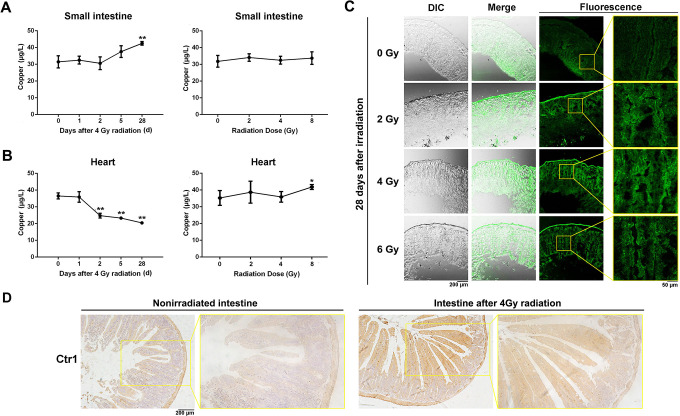
Ionizing radiation causes damage to a variety of tissues, especially radiation-sensitive tissues, such as the small intestine. Radiation-induced damage is caused primarily by increased oxidative stress in the body. Studies have shown that trace metal elements play an irreplaceable role in oxidative stress in humans, which may be associated with radiation-induced tissue damage. However, the alteration and functional significance of trace metal elements in radiation-induced injury is not clear. In this study, we explored the association between radiation-induced damage and 7 trace metal elements in mouse models. We found that the concentration of zinc and copper in mice serum was decreased significantly after irradiation, whereas that of nickel, manganese, vanadium, cobalt, and stannum was not changed by inductively coupled plasma mass spectrometry. The role of copper in radiation-induced intestines was characterized in detail. The concentration of copper was increased in irradiated intestine but reduced in irradiated heart. Immunohistochemistry staining showed that copper transporter protein copper transport 1 expression was upregulated in irradiated mouse intestine, suggesting its potential involvement in radiation-induced copper accumulation. At the cellular level, the addition of CuCl2 potentiated radiation-induced reactive oxygen species in intestine-derived human intestinal epithelial cell and IEC-6 cells. Moreover, the level of copper in damaged cells may be related to the severity of radiation-induced damage as evidenced by a cell viability assay. These results indicate that copper may be involved in the progression of radiation-induced tissue damage and may be a potential therapeutic target.

Keywords: copper; radiation; radiation-induced intestinal injury; trace metal element.


Conflict of interest statement

Declaration of Conflicting Interests: The author(s) declared no potential conflicts of interest with respect to the research, authorship, and/or publication of this article.

Figures

Figure 1.
Figure 1.
Changes of metal elements in serum at different time points after 4 Gy total body irradiation (TBI) in mice. C57BL/6N mice were randomized into 5 groups (n = 4) and irradiated with 4 Gy TBI using 250 kV X-rays. Serum samples were obtained 0, 1, 2, 5, or 28 days after irradiation. Inductively coupled plasma mass spectrometry was used to detect the metal concentration in serum. A, Serum zinc concentration. B, Serum copper concentration. C, Serum nickel concentration. D, Serum manganese concentration. E, Serum vanadium concentration. F, Serum cobalt concentration. G, Serum stannum concentration. *P < .05, **P < .01.
Figure 2.
Figure 2.
Changes of metal elements in serum after different radiation doses in mice. C57BL/6N mice were randomized into 4 groups (n = 4) and irradiated with a single dose of 0, 2, 4, and 8 Gy (total body irradiation) using 250 kV X-rays. Serum samples were obtained 24 hours after irradiation. Inductively coupled plasma mass spectrometry was used to detect the metal content in serum. A, Serum zinc concentration. B, Serum copper concentration. C, Serum nickel concentration. D, Serum manganese concentration. E, Serum vanadium concentration. F, Serum cobalt concentration. G, Serum stannum concentration. *P < .05, **P < .01.
Figure 3.
Figure 3.
Changes of copper levels in heart and small intestine tissues. A, The concentration of copper in small intestine tissue. Mice were either irradiated with 4 Gy X-rays and tissue samples were collected at different time points or irradiated with different doses and collected 24 hours after radiation. B, Changes in copper levels in mouse heart tissue. Mice were either irradiated with 4 Gy X-rays and tissue samples were collected at different time points or irradiated with different doses and collected 24 hours after radiation. C, Frozen sections were prepared from small intestine tissue 28 days after irradiation with different doses. Cu2+ in the sections was determined with a copper-specific probe (C27H29N5O2S). Sections were observed with a confocal microscope. *P < .05, **P < .01. D, Immunohistochemistry staining of Ctr1 in nonirradiated and 4 Gy irradiated mouse intestinal tissues.
Figure 4.
Figure 4.
Radiation induces Cu2+ accumulation in intestinal cells. Both (A) HIEC and (B) IEC-6 cells were treated with CuCl2 (40 μM) and irradiated with different doses. After 24 hours, the Cu2+ in the cells were stained with C27H29N5O2 S and observed by confocal microscopy. C, Quantitative analysis of Cu2+ accumulation in intestinal cells ImageJ software. *P < .05, **P < .01. HIEC indicates human intestinal epithelial cell; ns, nonsignificant.
Figure 5.
Figure 5.
Effect of Cu2+ on the radiation-induced ROS and cell viability after irradiation. A, Both HIEC and IEC-6 cells were treated with CuCl2 (40 μM) and irradiated with different doses, and cell ROS were observed with a fluorescence microscope at 24 and 48 hours. Both HIEC and IEC-6 cells were treated with different concentrations of CuCl2 and irradiated with different doses. Cell proliferation was measured with a CCK-8 cell viability assay (B) 24 and (C) 48 hours after radiation. CCK-8 indicates Cell Counting Kit-8; HIEC, human intestinal epithelial cell; ROS, reactive oxygen species.

References

    1. Imanaka T, Hayashi G, Endo S. Comparison of the accident process, radioactivity release and ground contamination between Chernobyl and Fukushima-1. J Radiat Res. 2015;56(suppl):i56–i61. - PMC - PubMed
    1. Burnet NG, Nyman J, Turesson I, Wurm R, Yarnold JR, Peacock JH. Prediction of normal-tissue tolerance to radiotherapy from in-vitro cellular radiation sensitivity. Lancet. 1992;339(8809):1570–1571. - PubMed
    1. Zhao Y, Zhang J, Han X, Fan S. Total body irradiation induced mouse small intestine senescence as a late effect. J Radiat Res. 2019;60(4):442–450. - PMC - PubMed
    1. Jomova K, Valko M, Advances in metal-induced oxidative stress and human disease. Toxicology. 2011;283(2-3):65–87. - PubMed
    1. Kambe T, Tsuji T, Hashimoto A, Itsumura N. The physiological, biochemical, and molecular roles of zinc transporters in zinc homeostasis and metabolism. Physiol Rev. 2015;95(3):749–784. - PubMed

LinkOut - more resources