

Improvements to the retractor and muscle flap design for minimally invasive cochlear implantation
- PMID: 32110240
- PMCID: PMC7033583
- DOI: 10.1016/j.joto.2019.09.003
Improvements to the retractor and muscle flap design for minimally invasive cochlear implantation
Erratum in
-
Erratum regarding missing Declaration of Competing Interest statements in previously published articles.J Otol. 2020 Dec;15(4):179. doi: 10.1016/j.joto.2020.09.006. Epub 2020 Sep 26. J Otol. 2020. PMID: 33293923 Free PMC article.
Abstract
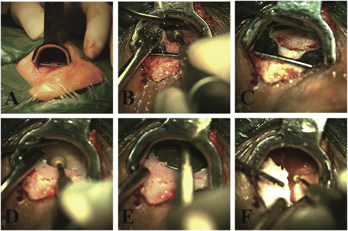
Objective: The aim of this study was to improve muscle flaps and to evaluate surgical outcomes with the use of a novel specialized retractor, which is a surgical instrument used to locate and shape a bony seat for minimally invasive cochlear implantation.
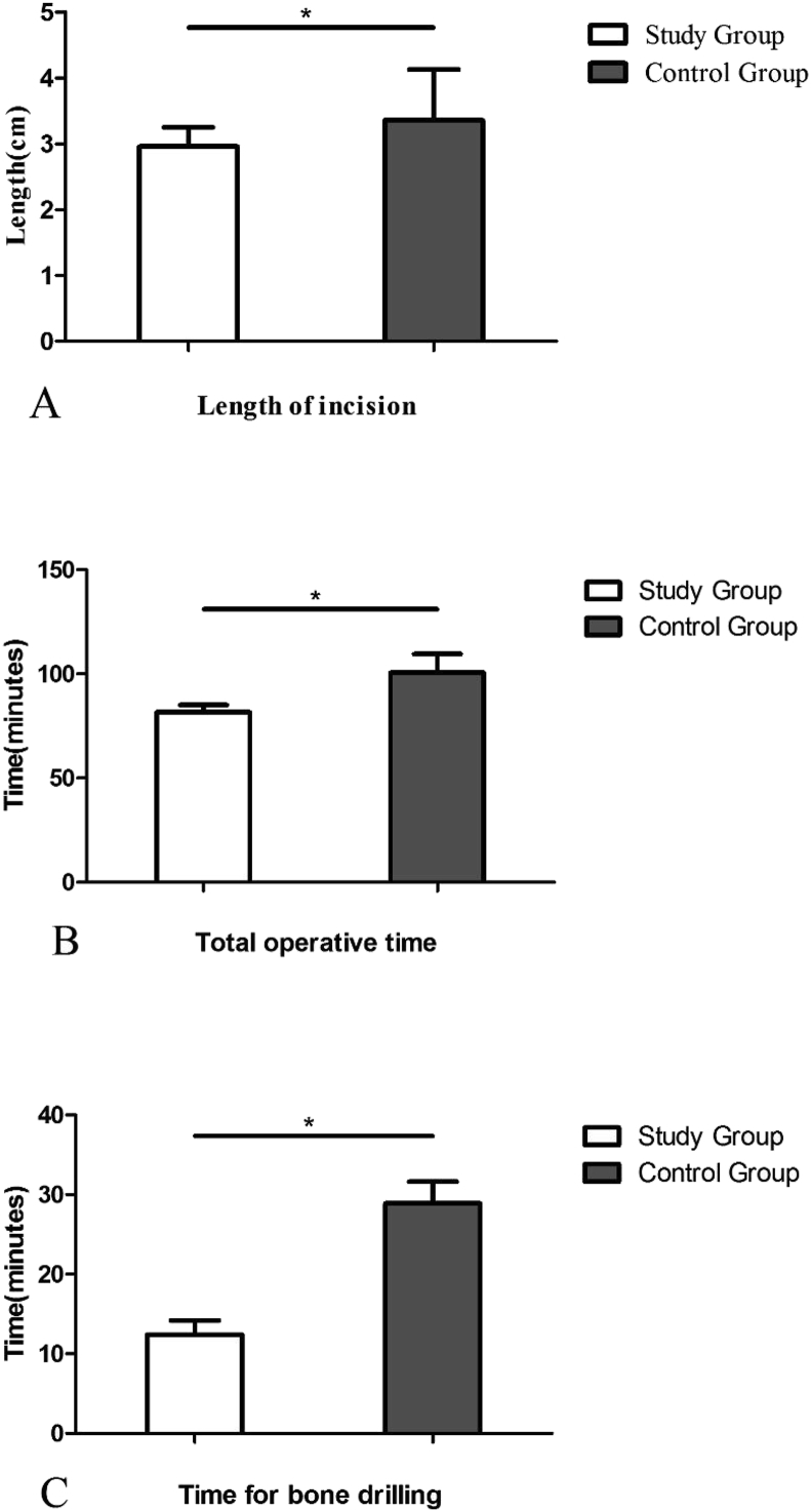
Methods: 50 patients aged 1-75 years with sensorineural hearing loss who required cochlear implantation were recruited. A small incision (<3 cm) was made, and the novel specialized retractor was used in the study group during cochlear implantation. The incision length, surgical outcomes and operative time were recorded and analyzed.
Results: The incision length, total operative time and drilling bony time were shorter in the study group than in the control group (P < 0.05, respectively). All patients recovered well after the surgery without any severe complications.
Conclusion: The use of a novel specialized retractor standardized the surgical processes of cochlear implantation. The retractor helped locate and control the size of the bony well during bone drilling. The tool reduced the technical difficulty and improved the efficacy of this minimally invasive operation.
Keywords: Cochlear implantation; Minimally invasive surgery; Muscle flap; Retractor.
© 2020 PLA General Hospital Department of Otolaryngology Head and Neck Surgery. Production and hosting by Elsevier (Singapore) Pte Ltd.
Figures


References
-
- Bajaj Y., Wyatt M., Hartley B. Small postaural incision for paediatric cochlear implantation. Cochlear Implants Int. 2005;6(2):77–84. - PubMed
-
- Balkany T.J., Whitley M., Shapira Y. The temporalis pocket technique for cochlear implantation: an anatomic and clinical study. Otol. Neurotol. 2009;30(7):903–907. - PubMed
-
- Caversaccio M., Gavaghan K., Wimmer W. Robotic cochlear implantation: surgical procedure and first clinical experience. Acta Otolaryngol. 2017;137(4):447–454. - PubMed
-
- Dalchow C.V., Werner J.A. A new instrument for minimal access surgery in cochlear implantation. Otol. Neurotol. 2005;26(4):678–679. - PubMed
LinkOut - more resources
Full Text Sources
