. 2019 Oct 3;10(45):10595-10600.
doi: 10.1039/c9sc04153b. eCollection 2019 Dec 7.

Twists or turns: stabilising alpha vs. beta turns in tetrapeptides

Affiliations

Twists or turns: stabilising alpha vs. beta turns in tetrapeptides

Huy N Hoang et al. Chem Sci. .

Abstract

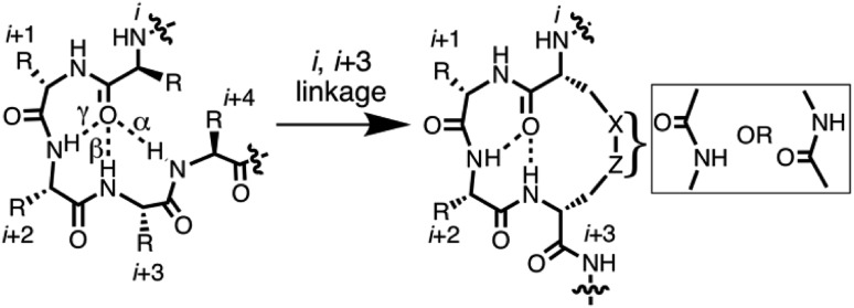
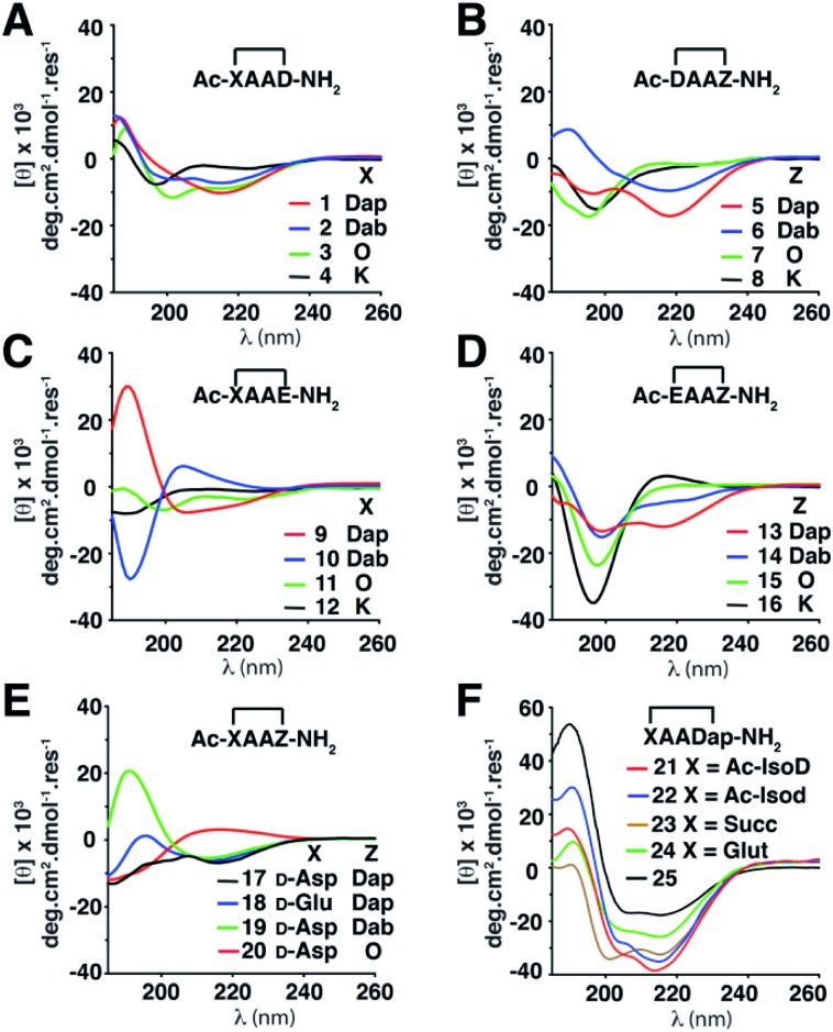
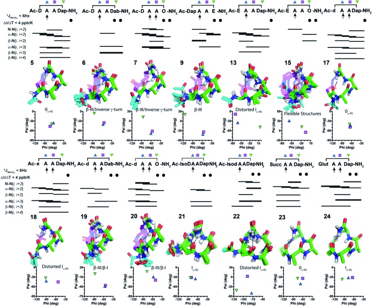
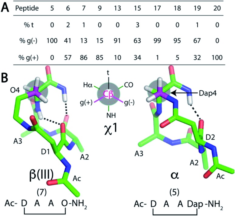
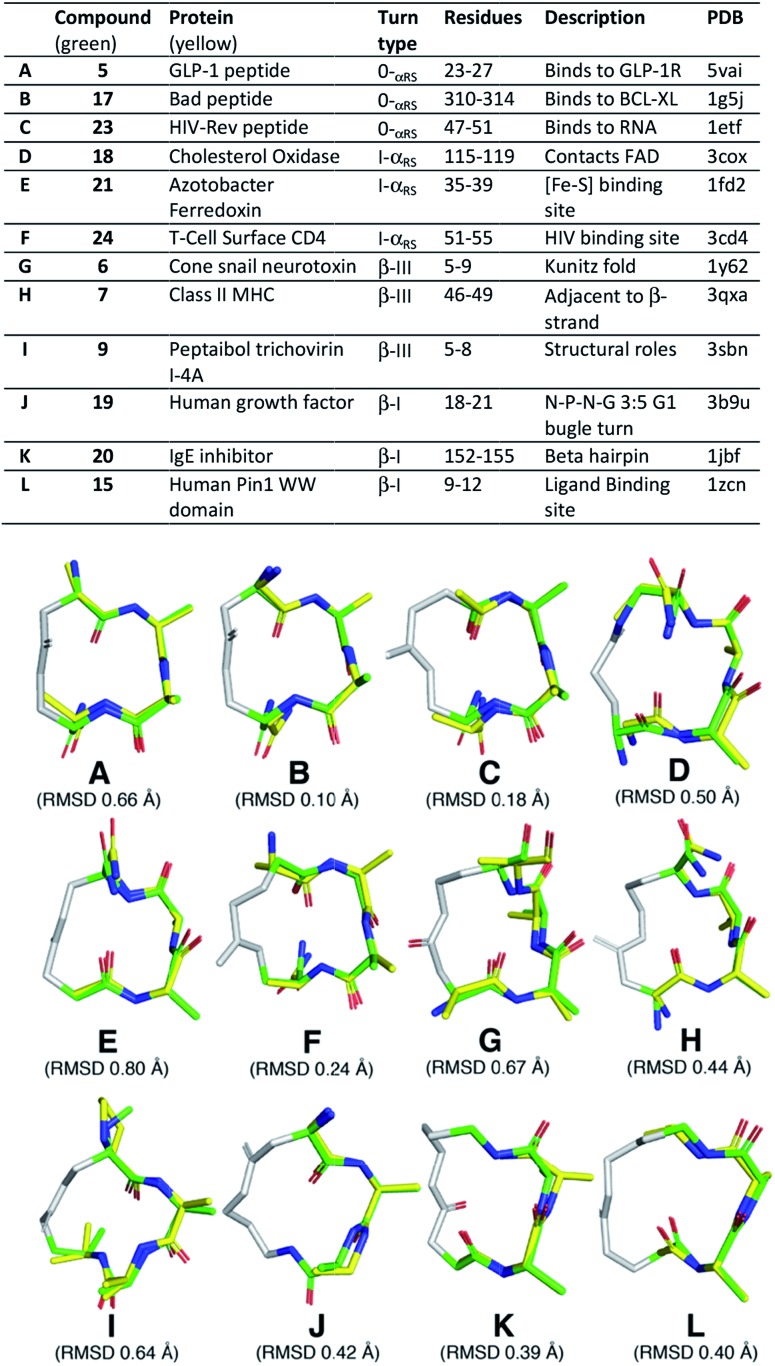
Protein-protein interactions involve hotspots as small as 4 sequential amino acids. Corresponding tetrapeptides have no structure in water. Here we report linking side chains of amino acids X and Z to form 24 cyclic tetrapeptides, cyclo-[XAAZ]-NH2, and stabilise 14-18 membered rings that mimic different kinds of non-regular secondary structures found in protein hotspots. 2D NMR spectra allowed determination of 3D structures for 14 cyclic tetrapeptides in water. Five formed two (i, i + 3) hydrogen bonds and a beta/gamma (6, 7) or beta (9, 19, 20) turn; eight formed one (i, i + 4) hydrogen bond and twisted into a non-helical (13, 18, 21, 22, 24) or helical (5, 17, 23) alpha turn; one was less structured (15). A beta or gamma turn was favoured for Z = Dab, Orn or Glu due to a χ1 gauche (+) rotamer, while an alpha turn was favoured for Z = Dap (but not X = Dap) due to a gauche (-) rotamer. Surprisingly, an unstructured peptide ARLARLARL could be twisted into a helix when either a helical or non-helical alpha turn (5, 13, 17, 18, 21-24) with Z = Dap was attached to the N-terminus. These structural models provide insights into stability for different turns and twists corresponding to non-regular folds in protein hotspots.


Figures

Fig. 1
Fig. 1. 7-, 10-, 13-membered hydrogen bonded rings define γ-, β- and α-turns respectively (left) that potentially might be stabilised by cyclisation via side chains (right).
Fig. 2
Fig. 2. CD spectra of cyclic peptides (150 μM) in aqueous phosphate buffer (10 mM, pH 7.4, 25 °C). (A) 1–4. (B) 5–8. (C) 9–12. (D) 13–16. (E) 17–20. (F) 21–25. Standard 1-letter codes for l-amino acids unless otherwise indicated. Dap = l-2,3-diaminopropionic acid; Dab = l-2,4-diaminobutyric acid; O = l-ornithine; Succ = succinic acid; Glut = glutaric acid; IsoD and Isod are l- and d-aspartic acid respectively, where a side chain carbonyl instead forms the amide bond with the backbone nitrogen atom of the next residue.
Fig. 3
Fig. 3. NMR summaries, 20 lowest energy structures, Ramachandran plots and turn types in water for fourteen cyclic tetrapeptides: 5–7, 9, 13, 15, 17–24 in H2O/D2O (9 : 1) at 298 K. Compounds 5, 13, 17, 18, 21–24 form one hydrogen bond; while 6, 7, 9, 15, 19, 20 form two hydrogen bonds.
Fig. 4
Fig. 4. (A) Rotamer populations for residue Z4 from 50 ns MD simulations of cyclic peptides 5–20. NMR structures of: (B) 7 and 5 show how the χ1 angle of residue Z4 controls β(III)-turn vs. α-turn structures.
Fig. 5
Fig. 5. Average backbone solution structures of 5–7, 9, 15, 17–21 and 23–24 (green; residues: A, A, X/Z) superimposed on the three central, consecutive, residues of an α-helix, α-turn, β-III turn and β-I turn from protein crystal structures (yellow). The linkers in these compounds are coloured grey.

References

    1. Hutchinson E. G., Thornton J. M. Protein Sci. 1994;3:2207–2216. - PMC - PubMed
    2. Wilmot C. M., Thornton J. M. Protein Eng. 1990;3:479–493. - PubMed
    1. Craik D. J., Fairlie D. P., Liras S., Price D. Chem. Biol. Drug Des. 2013;81:136–147. - PubMed
    2. Nevola L., Giralt E. Chem. Commun. 2015;51:3302–3315. - PubMed
    3. Tyndall J. D., Pfeiffer B., Abbenante G., Fairlie D. P. Chem. Rev. 2005;105:793–826. - PubMed
    1. Harrison R. S., PhD dissertation, University of Queensland, 2011.
    2. Bergey C. M., Watkins A. M., Arora P. S. Bioinformatics. 2013;29:2806–2807. - PMC - PubMed
    3. Gavenonis J., Sheneman B. A., Siegert T. R., Eshelman M. R., Kritzer J. A. Nat. Chem. Biol. 2014;10:716–722. - PMC - PubMed
    1. Pavone V., Gaeta G., Lombardi A., Nastri F., Maglio O., Isernia C., Saviano M. Biopolymers. 1996;38:705–721. - PubMed
    2. Nataraj D. V., Srinivasan N., Sowdhamini R., Ramakrishnan C. Curr. Sci. 1995;69:434–447.
    1. Hoang H. N., Driver R. W., Beyer R. L., Malde A. K., Le G. T., Abbenante G., Mark A. E., Fairlie D. P. Angew. Chem., Int. Ed. 2011;50:11107–11111. - PubMed