

Long-term study on the osteogenetic capability and mechanical behavior of a new resorbable biocomposite anchor in a canine model
- PMID: 32110507
- PMCID: PMC7033359
- DOI: 10.1016/j.jot.2019.12.008
Long-term study on the osteogenetic capability and mechanical behavior of a new resorbable biocomposite anchor in a canine model
Abstract
Background: Biodegradable suture anchors are commonly used for repairing torn rotator cuffs, but these biodegradable materials still suffer from low mechanical strength, poor osteointegration, and the generation of acidic degradation byproducts.
Method: The purpose of this study was to evaluate the long-term mechanical behavior and osteogenetic capabilities of a biocomposite anchor injection molded with 30% β-tricalcium phosphate microparticles blended with 70% poly (L-lactide-co-glycolide) (85/15). This study investigated in vitro degradation and in vivo bone formation in a canine model. The initial mechanical behavior, mechanical strength retention with degradation time, and degradation features were investigated.
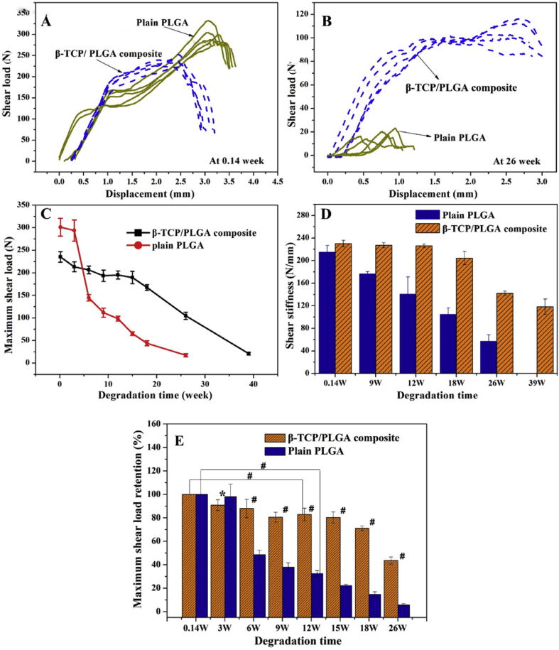
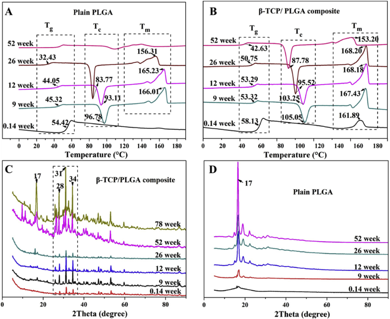
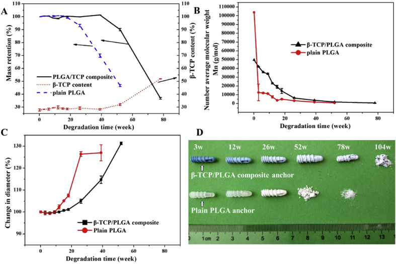
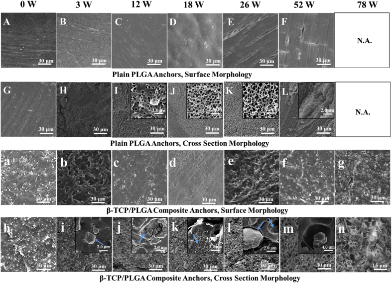
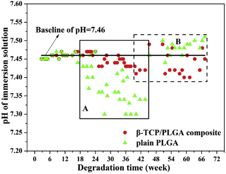
Results: The results showed that the biocomposite anchor had sufficient initial mechanical stability confirmed by comparing the initial shear load on the anchor with the minimum shear load borne by an ankle fracture fixation screw, which is considered a worst-case implantation site for mechanical loading. The maximum shear load retention of the biocomposite anchor was 83% at 12 weeks, which is desirable, as it aligns with the rate of bone healing. The β-tricalcium phosphate fillers were evenly dispersed in the polymeric matrix and acted to slow the degradation rate and improve the mechanical strength of the anchor. The interface characteristics between the β-tricalcium phosphate particles and the polymeric matrix changed the degradation behavior of the biocomposite. Phosphate buffer saline was shown to diffuse through the interface into the biocomposite to inhibit the core accelerated degradation rate. In vivo, the addition of β-tricalcium phosphate induced new bone formation. The biocomposite material developed in this study demonstrated improved osteogenesis in comparison to a plain poly (L-lactide-co-glycolide) material. Neither anchor produced adverse tissue reactions, indicating that the biocomposite had favorable biocompatibility following long-term implantation.
Conclusion: In summary, the new biocomposite anchor presented in this study had favorable osteogenetic capability, mechanical property, and controlled degradation rate for bone fixation.
Translational potential of this article: The new biocomposite anchor had sufficient initial and long-term fixation stability and bone formation capability in the canine model. It is indicated that the new biocomposite anchor has a potential for orthopedic application.
Keywords: Anchor-bone interaction; Biocomposite anchor; Degradation rate; Mechanical property; Osteogenesis.
© 2020 The Authors.
Conflict of interest statement
The authors have no conflicts of interest to disclose in relation to this article.
Figures


Similar articles
-
The degradation outcome of biocomposite suture anchors made from poly L-lactide-co-glycolide and β-tricalcium phosphate.Arthroscopy. 2013 Nov;29(11):1834-9. doi: 10.1016/j.arthro.2013.08.004. Arthroscopy. 2013. PMID: 24209681
-
Degradation of Cylindrical Poly-Lactic Co-Glycolide/Beta-Tricalcium Phosphate Biocomposite Anchors After Arthroscopic Bankart Repair: A Prospective Study.Orthopedics. 2018 May 1;41(3):e348-e353. doi: 10.3928/01477447-20180226-08. Epub 2018 Mar 2. Orthopedics. 2018. PMID: 29494743
-
Long-term degradation of a poly-lactide co-glycolide/β-tricalcium phosphate biocomposite interference screw.Arthroscopy. 2011 May;27(5):637-43. doi: 10.1016/j.arthro.2010.11.056. Epub 2011 Mar 23. Arthroscopy. 2011. PMID: 21429700 Clinical Trial.
-
Perianchor Cyst Formation Around Biocomposite Biodegradable Suture Anchors After Rotator Cuff Repair.Am J Sports Med. 2015 Dec;43(12):2907-12. doi: 10.1177/0363546515608484. Epub 2015 Oct 19. Am J Sports Med. 2015. PMID: 26482545
-
The formation of perianchor fluid associated with various suture anchors used in rotator cuff repair: all-suture, polyetheretherketone, and biocomposite anchors.Bone Joint J. 2019 Dec;101-B(12):1506-1511. doi: 10.1302/0301-620X.101B12.BJJ-2019-0462.R2. Bone Joint J. 2019. PMID: 31786997 Clinical Trial.
Cited by
-
Comparison between all-suture and biocomposite anchors in the arthroscopic treatment of traumatic anterior shoulder instability: A retrospective cohort study.J Orthop. 2021 Mar 27;24:264-270. doi: 10.1016/j.jor.2021.03.011. eCollection 2021 Mar-Apr. J Orthop. 2021. PMID: 33867751 Free PMC article.
-
An overview of polyester/hydroxyapatite composites for bone tissue repairing.J Orthop Translat. 2021 Apr 1;28:118-130. doi: 10.1016/j.jot.2021.02.005. eCollection 2021 May. J Orthop Translat. 2021. PMID: 33898248 Free PMC article.
References
-
- Chaudhry S., Dehne K., Hussain F. A review of suture anchors. Orthop Traumatol. 2019;33(4):263–270.
-
- Bishop J., Klepps S., Lo I.K., Bird J., Gladstone J.N., Flatow E.L. Cuff integrity after arthroscopic versus open rotator cuff repair: a prospective study. J Shoulder Elb Surg. 2006;15(3):290–299. - PubMed
-
- Ricchetti E.T., Aurora A., Iannotti J.P., Derwin K.A. Scaffold devices for rotator cuff repair. J Shoulder Elb Surg. 2012;21(2):251–265. - PubMed
LinkOut - more resources
Full Text Sources
Research Materials
