

Intravascular T-cell lymphoma in a patas monkey (Erythrocebus patas)
- PMID: 32110691
- PMCID: PMC7041513
- DOI: 10.5194/pb-4-39-2017
Intravascular T-cell lymphoma in a patas monkey (Erythrocebus patas)
Abstract
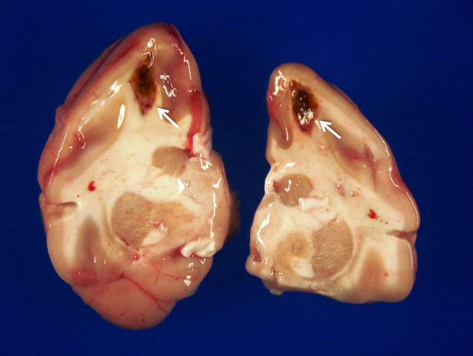
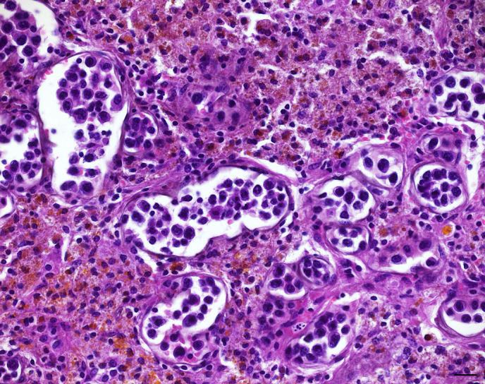
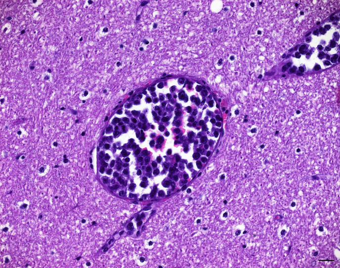
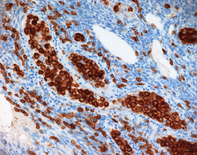
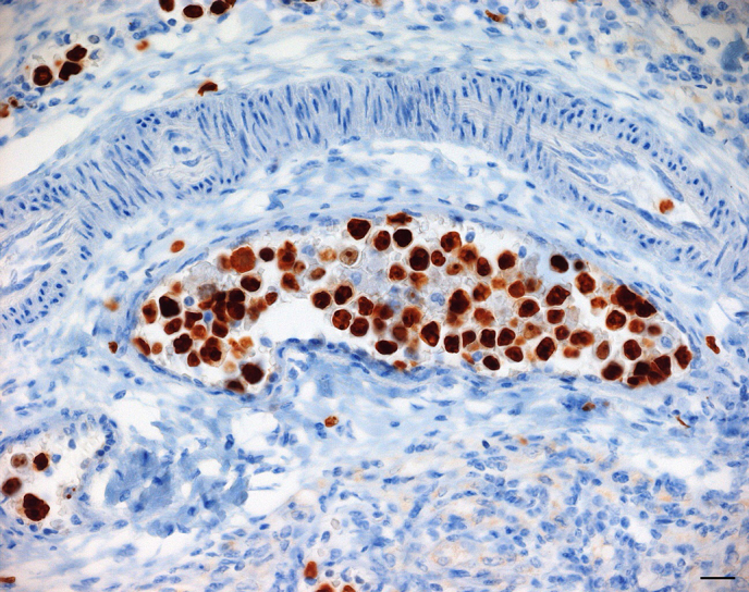
A 9-year-old female captive patas monkey (Erythrocebus patas) presented with poor general condition, inability to stand, petechiae, anaemia, thrombocytopenia, and leukocytosis. Due to poor response to treatment, the animal was euthanized 16 days later. Postmortem examination revealed hemorrhages in several organs and bilateral cerebral infarctions. Histologically, prominent accumulations of large neoplastic lymphocytes in cerebral and meningeal blood vessels were demonstrated within the lesions and in other organs (e.g., bone marrow, ovary, intestine). Immunohistochemically, neoplastic cells expressed CD3 and Ki-67. PCR revealed a lymphocryptovirus (LCV) infection, while Epstein-Barr nuclear antigen 2 (EBNA2) could not be demonstrated within neoplastic cells by means of immunohistochemistry. Based on the pathological findings, an intravascular lymphoma (IVL) of T-cell origin was diagnosed. To the authors' knowledge, this is the first report on this rare entity in a nonhuman primate.
Copyright: © 2017 Karen Lampe et al.
Conflict of interest statement
The authors declare that they have no conflict of interest.
Figures
References
-
- Allan JS, Leland M, Broussard S, Mone J, Hubbard G. Simian T-cell lymphotropic viruses (STLVs) and lymphomas in African nonhuman primates. Cancer Invest. 2001;19:383–395. - PubMed
-
- Au WY, Shek WH, Nicholls J, Tse KM, Todd D, Kwong YL. T-cell intravascular lymphomatosis (angiotropic large cell lymphoma): Association with Epstein-Barr viral infection. Histopathology. 1997;31:563–567. - PubMed
-
- Bibollet-Ruche F, Galat-Luong A, Cuny G, Sarni-Manchado P, Galat G, Durand JP, Pourrut X, Veas F. Simian immunodeficiency virus infection in a patas monkey (Erythrocebus patas): evidence for cross-species transmission from African green monkeys (Cercopithecus aethiops sabaeus) in the wild. J Gen Virol. 1996;77:773–781. - PubMed
-
- Blaschke S, Hannig H, Buske C, Kaup FJ, Hunsmann G, Bodemer W. Expression of the simian Epstein-Barr virus-encoded latent membrane protein-1 in malignant lymphomas of SIV-infected rhesus macaques. J Med Virol. 2001;65:114–120. - PubMed
LinkOut - more resources
Full Text Sources