

Spontaneous meningioma in a pig-tailed macaque (Macaca nemestrina)
- PMID: 32110712
- PMCID: PMC7041523
- DOI: 10.5194/pb-5-7-2018
Spontaneous meningioma in a pig-tailed macaque (Macaca nemestrina)
Abstract
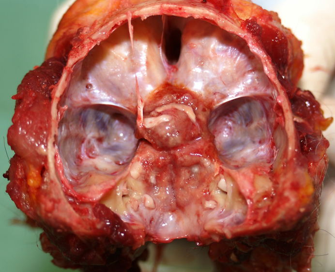
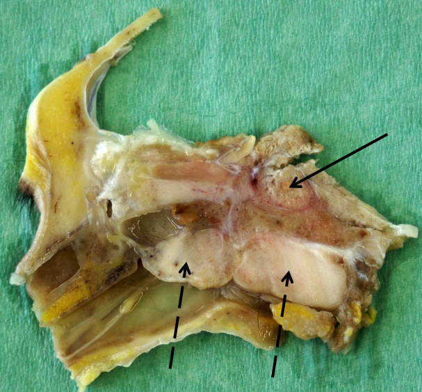
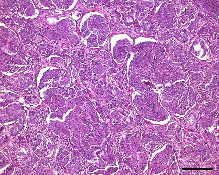
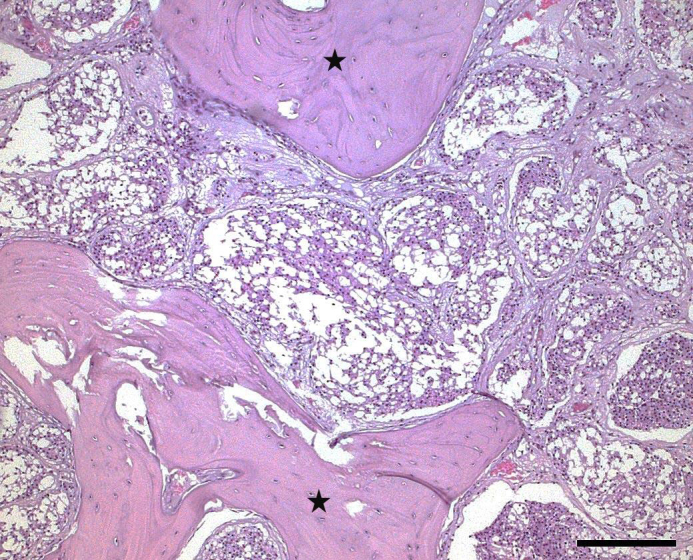
We present a case of spontaneous meningioma in a female pig-tailed macaque (Macaca nemestrina) more than 24 years old. Clinically, the monkey displayed slow, weak, and insecure movements and poor vision. A tumorous mass was present at the floor of the cranial vault extending from the optic chiasm towards the foramen magnum. It compressed adjacent parts of the brain, infiltrated the sphenoidal and occipital bone, and showed transcranial expansion into the pharyngeal area. Histologically, the tumor was consistent with a meningioma displaying mostly meningothelial and some microcystic components. Since only six cases of meningiomas in nonhuman primates have been reported so far and only two of these meningiomas have been described in detail, the findings of each case should be reported to expand the knowledge base of this type of tumor. In addition, this is the first description of a meningioma in pig-tailed macaques.
Copyright: © 2018 Roland Plesker et al.
Conflict of interest statement
The authors declare that they have no conflict of interest.
Figures
Similar articles
-
Taxonomy, Evolutionary and Dispersal Events of Pig-Tailed Macaque, Macaca nemestrina (Linnaeus, 1766) in Southeast Asia with Description of a New Subspecies, Macaca nemestrina perakensis in Malaysia.Zool Stud. 2021 Oct 8;60:e50. doi: 10.6620/ZS.2021.60-50. eCollection 2021. Zool Stud. 2021. PMID: 35003344 Free PMC article.
-
Blood groups of pig-tailed macaques (Macaca nemestrina).Am J Phys Anthropol. 1978 Mar;48(3):321-30. doi: 10.1002/ajpa.1330480308. Am J Phys Anthropol. 1978. PMID: 416724
-
An outbreak of tuberculosis in endangered northern pig-tailed macaques (Macaca leonina) and milu deer (Elaphurus davidianus) from a zoo in China.Vet Med Sci. 2023 Mar;9(2):992-998. doi: 10.1002/vms3.1014. Epub 2023 Jan 10. Vet Med Sci. 2023. PMID: 36626281 Free PMC article.
-
[Measurement and analysis of hematology and blood chemistry parameters in northern pig-tailed macaques (Macaca leonina)].Dongwuxue Yanjiu. 2013 Apr;34(2):89-96. doi: 10.3724/SP.J.1141.2013.02089. Dongwuxue Yanjiu. 2013. PMID: 23572357 Review. Chinese.
-
Spinal Accessory Nerve Meningioma at the Foramen Magnum with Medullar Compression: A Case Report and Literature Review.World Neurosurg. 2019 Aug;128:158-161. doi: 10.1016/j.wneu.2019.05.013. Epub 2019 May 10. World Neurosurg. 2019. PMID: 31082561 Review.
Cited by
-
Multiple adenomas of the thyroid gland in an African green monkey (Chlorocebus aethiops).Primate Biol. 2023 May 12;10(1):1-6. doi: 10.5194/pb-10-1-2023. eCollection 2023. Primate Biol. 2023. PMID: 39149141 Free PMC article.
References
-
- Cantile C, Youssef S. Nervous System. In: Maxie MG, editor. Jubb, Kennedy and Palmer's Pathology of Domestic Animals. 6th ed. Elsevier; St. Louis, USA: 2016. pp. 251–406.
-
- Dahme E. Meningiome bei Fleischfressern. Berliner und Münchener Tierärztliche Wochenschrift. 1957;70:32–34.
-
- Devaprasath A, Chack G. Diagnostic validity of the Ki-67 labelling index using the MIB-1 monoclonal antibody in the grading of meningioma. Neurol India. 2003;51:336–340. - PubMed
-
- Enam SA, Abdulrauf S, Metha GM, Mahmood A. Metastasis in meningioma. Acta Neuropathol. 2005;138:1172–1178. - PubMed
LinkOut - more resources
Full Text Sources