

Microblotches on Dermoscopy of Melanocytic Lesions are Associated with Melanoma: A Cross-sectional Study
- PMID: 32110813
- PMCID: PMC9128959
- DOI: 10.2340/00015555-3436
Microblotches on Dermoscopy of Melanocytic Lesions are Associated with Melanoma: A Cross-sectional Study
Abstract
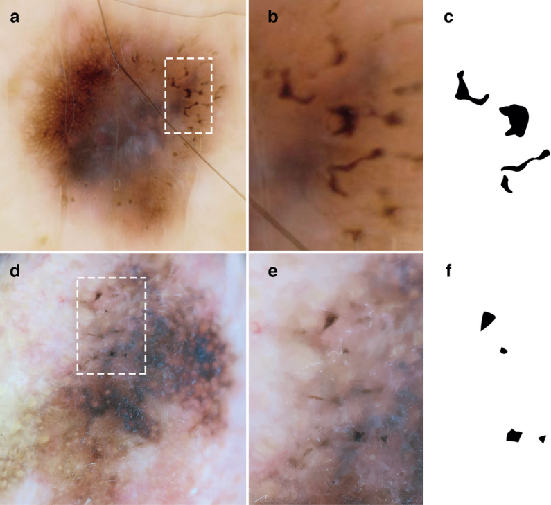
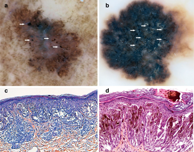
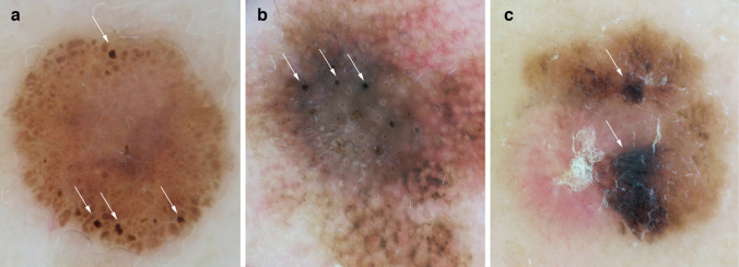
Numerous dermoscopic structures for the early detection of melanoma have been described. The aim of this study was to illustrate the characteristics of dermoscopic structures that are similar to blotches, but smaller (termed microblotches), and to evaluate their association with other well-known dermoscopic structures. A cross-sectional study design, including 165 dermoscopic images of melanoma was used to define microblotches, and 241 consecutive images of naevi from the HAM10000 database, were studied to evaluate the prevalence of this criterion in both groups. Microblotches were defined as sharply demarcated structures ≤1 mm, with geographical borders visible only with dermoscopy. Microblotches were present in 38.7% of the melanomas and 6.7% of the naevi. Moreover, microblotches were associated with an odds ratio (OR) of malignancy of 5.79, and were more frequent in invasive melanoma than in the in-situ subtype (OR 2.92). Histologically, they correspond to hyperpigmented parakeratosis or epidermal consumption. In conclusion, microblotches are related to melanomas. This finding could help dermatologists to differentiate between naevi and melanomas.
Keywords: dermatoscopy; melanoma; microblotches; prognosis; dermoscopy.
Conflict of interest statement
Figures
References
-
- van der Rhee JI, Bergman W, Kukutsch NA. The impact of dermoscopy on the management of pigmented lesions in everyday clinical practice of general dermatologists: a prospective study. Br J Dermatol 2010; 162: 563–567. - PubMed
-
- Vestergaard ME, Macaskill P, Holt PE, Menzies SW. Dermoscopy compared with naked eye examination for the diagnosis of primary melanoma: a meta-analysis of studies performed in a clinical setting. Br J Dermatol 2008; 159: 669–676. - PubMed
-
- Carli P, de Giorgi V, Chiarugi A, Nardini P, Weinstock MA, Crocetti E, et al. Addition of dermoscopy to conventional naked-eye examination in melanoma screening: a randomized study. J Am Acad Dermatol 2004; 50: 683–689. - PubMed
-
- González-Álvarez T, Carrera C, Bennassar A, Vilalta A, Rull R, Alós L, et al. Dermoscopy structures as predictors of sentinel lymph node positivity in cutaneous melanoma. Br J Dermatol 2015; 172: 1269–1277. - PubMed
-
- Ribero S, Argenziano G, Lallas A, Moscarella E, Benati E, Raucci M, et al. Dermoscopic features predicting the presence of mitoses in thin melanoma. J Dermatol Sci 2017; 86: 158–161. - PubMed
