Review
. 2020 Feb 26;9(3):540.
doi: 10.3390/cells9030540.

Translational Regulations in Response to Endoplasmic Reticulum Stress in Cancers

Affiliations
Review

Translational Regulations in Response to Endoplasmic Reticulum Stress in Cancers

Manon Jaud et al. Cells. .

Abstract

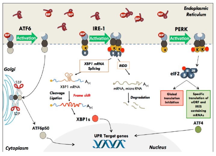
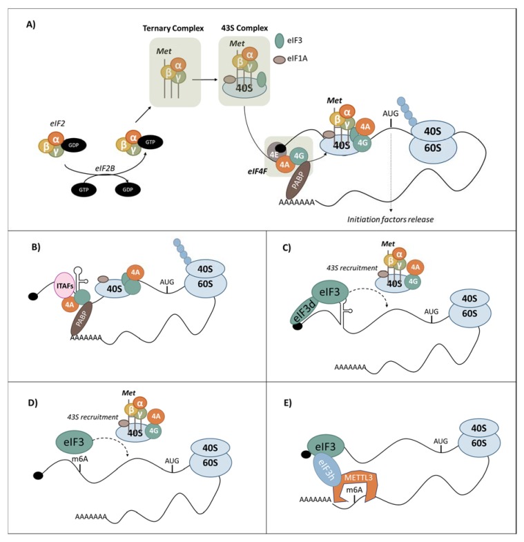
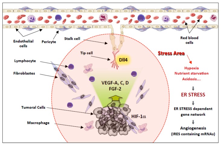
During carcinogenesis, almost all the biological processes are modified in one way or another. Among these biological processes affected, anomalies in protein synthesis are common in cancers. Indeed, cancer cells are subjected to a wide range of stresses, which include physical injuries, hypoxia, nutrient starvation, as well as mitotic, oxidative or genotoxic stresses. All of these stresses will cause the accumulation of unfolded proteins in the Endoplasmic Reticulum (ER), which is a major organelle that is involved in protein synthesis, preservation of cellular homeostasis, and adaptation to unfavourable environment. The accumulation of unfolded proteins in the endoplasmic reticulum causes stress triggering an unfolded protein response in order to promote cell survival or to induce apoptosis in case of chronic stress. Transcription and also translational reprogramming are tightly controlled during the unfolded protein response to ensure selective gene expression. The majority of stresses, including ER stress, induce firstly a decrease in global protein synthesis accompanied by the induction of alternative mechanisms for initiating the translation of mRNA, later followed by a translational recovery. After a presentation of ER stress and the UPR response, we will briefly present the different modes of translation initiation, then address the specific translational regulatory mechanisms acting during reticulum stress in cancers and highlight the importance of translational control by ER stress in tumours.

Keywords: ER stress; translation initiation; uORF; unfolded protein response (UPR), IRES.


Conflict of interest statement

The authors declare no conflict of interest.

Figures

Figure 1
Figure 1
The different UPR effectors and their modes of action. In the basal state, the three UPR effector transmembrane proteins (PERK, ATF6, and IRE-1) are maintained inactive through their interaction with the protein chaperone BiP. The accumulation of poorly folded polypeptides in the ER lumen results in dissociation of BiP and activation of UPR. –I- PERK dimerises and phosphorylates the eIF2α subunit, leading to a global translation initiation inhibition. Specific mRNA subsets, containing cis-acting elements in their 5′UTR, such as uORF and IRES, escape translational inhibition triggered by eIF2 phosphorylation. –II- IRE-1 initiates an unconventional splicing of XBP-1 mRNA, as well as the degradation of some RNAs (this mechanism has been called RIDD for Regulated Ire1-Dependent Decay) –III- ATF6 traffics to the Golgi where proteolysis liberates its transcription factor amino-terminal domain, which is nuclearised and activates the expression of target genes.
Figure 2
Figure 2
Currently known processes of translation initiation (A) Cap-dependent mechanism of translation. The eukaryotic initiation factor 2 (eIF2)-GDP is recycled in eIF2-GTP by the enzyme eIF2B. eIF2-GTP binds the methionine transfer RNA (Met-tRNAi) in order to form the ternary complex which integrates the 43S complex comprising the 40S ribosome subunit, eukaryotic initiation factors (eIF3, eIF1/1A) and the ternary complex. 43S is recruited to the mRNA through the m7G cap by interacting with the eIF4F complex (eIF4E, eIF4A, eIF4G) and 43S scans the 5′UTR until the first starting codon. The codon/anti-codon interaction triggers the release of initiation factors and the recruitment of the 60S, and then elongation can start. (B) Internal ribosome entry sites (IRES)-mediated translation initiation. The IRES directly recruits ribosomes, thereby bypassing the requirement of the mRNA 5′ cap structure. (C) The binding of the cap by the eIF3d subunit in presence of the stem-loop in the 5′ UTR can bypass the canonical eIF4E translation and initiate an eIF3d-directed cap-dependent mRNA translation. (D) A single 5′ UTR-located N(6)-methyladenosine m(6)A can promote cap-independent mRNA translation initiation, through direct interaction with eIF3 which is sufficient to recruit the 43S complex and initiate translation even in the absence of the cap-binding factor eIF4E. (E) METTL3 enhances translation of mRNA containing m(6)A in its 3′UTR through interaction with eIF3h.
Figure 3
Figure 3
Schematic model of the network of gene expression co-regulation by IRES elements in stress conditions during tumoural progression. During tumour progression, the stress zone encompasses the growing tumour, but also its microenvironment. Both the tumour and the neo vessels, more particularly the Tip cells located at their extremity, which guide the neo vessels towards the tumour, are located in this unfavourable microenvironment. Hypoxia, nutrient starvation, and acidosis will irremediably induce the accumulation of unfolded protein in the reticulum of cells located in this area, leading to endoplasmic reticulum stress and UPR activation. Thus, in addition to transcriptional regulations, the activation of the PERK pathway will induce the co-regulation an UPR-dependent gene network containing IRES elements, revealing a translational regulon in which the synthesis of a cohort of angiogenic master regulator genes including VEGF-A,C,D, FGF-2, DLL4, and HIF1 is activated in response to ER stress. The fine-tuning of gene expression allows for efficient angiogenesis, which is a highly regulated process.

References

    1. Reid D.W., Nicchitta C.V. Diversity and selectivity in mRNA translation on the endoplasmic reticulum. Nat. Rev. Mol. Cell Biol. 2015;16:221–231. doi: 10.1038/nrm3958. - DOI - PMC - PubMed
    1. Ron D., Walter P. Signal integration in the endoplasmic reticulum unfolded protein response. Nat. Rev. Mol. Cell Biol. 2007;8:519–529. doi: 10.1038/nrm2199. - DOI - PubMed
    1. Hetz C. The unfolded protein response: Controlling cell fate decisions under ER stress and beyond. Nat. Rev. Mol. Cell Biol. 2012;13:89–102. doi: 10.1038/nrm3270. - DOI - PubMed
    1. Dejeans N., Manie S., Hetz C., Bard F., Hupp T., Agostinis P., Samali A., Chevet E. Addicted to secrete —Novel concepts and targets in cancer therapy. Trends Mol. Med. 2014;20:242–250. doi: 10.1016/j.molmed.2013.12.003. - DOI - PubMed
    1. Khan M.M., Nomura T., Chiba T., Tanaka K., Yoshida H., Mori K., Ishii S. The fusion oncoprotein PML-RARalpha induces endoplasmic reticulum (ER)-associated degradation of N-CoR and ER stress. J. Biol. Chem. 2004;279:11814–11824. doi: 10.1074/jbc.M312121200. - DOI - PubMed

Publication types