. 2020 Feb 26;9(3):539.
doi: 10.3390/cells9030539.

Conformational Switching in Bcl-xL: Enabling Non-Canonic Inhibition of Apoptosis Involves Multiple Intermediates and Lipid Interactions

Affiliations

Conformational Switching in Bcl-xL: Enabling Non-Canonic Inhibition of Apoptosis Involves Multiple Intermediates and Lipid Interactions

Victor Vasquez-Montes et al. Cells. .

Abstract

The inhibition of mitochondrial permeabilization by the anti-apoptotic protein Bcl-xL is crucial for cell survival and homeostasis. Its inhibitory role requires the partitioning of Bcl-xL to the mitochondrial outer membrane from an inactive state in the cytosol, leading to its extensive refolding. The molecular mechanisms behind these events and the resulting conformations in the bilayer are unclear, and different models have been proposed to explain them. In the most recently proposed non-canonical model, the active form of Bcl-xL employs its N-terminal BH4 helix to bind and block its pro-apoptotic target. Here, we used a combination of various spectroscopic techniques to study the release of the BH4 helix (α1) during the membrane insertion of Bcl-xL. This refolding was characterized by a gradual increase in helicity due to the lipid-dependent partitioning-coupled folding and formation of new helix αX (presumably in the originally disordered loop between helices α1 and α2). Notably, a comparison of various fluorescence and circular dichroism measurements suggested the presence of multiple Bcl-xL conformations in the bilayer. This conclusion was explicitly confirmed by single-molecule measurements of Fӧrster Resonance Energy Transfer from Alexa-Fluor-488-labeled Bcl-xL D189C to a mCherry fluorescent protein attached at the N-terminus. These measurements clearly indicated that the refolding of Bcl-xL in the bilayer is not a two-state transition and involves multiple membranous intermediates of variable compactness.

Keywords: BH4 domain; Bcl-2 proteins; Fluorescence Correlation Spectroscopy (FCS); Fluorescence Spectroscopy; Single Molecule FRET; apoptotic regulation; conformational switching; protein-membrane interactions.


Conflict of interest statement

The authors declare no conflict of interest.

Figures

Figure 1
Figure 1
Conformational switching of Bcl-xL in membranes, resulting in conversion from canonical to non-canonical forms of apoptotic inhibition. (a) The anti-apoptotic protein Bcl-xL (purple) binds to the pore former BAX (green) to block the permeabilization of the mitochondrial outer membrane (MOM) and prevent apoptosis [3,4]. (b) Several molecular mechanisms, involving both membrane-anchored and membrane-inserted Bcl-xL, have been proposed to explain this process. The canonical mode (left) relies on the interaction of anchored Bcl-xL with the BH3 helix of BAX [7]; while in the non-canonical mode (right), BAX binds the N-terminal BH4 helix of refolded and inserted Bcl-xL. Lipid composition is hypothesized to modulate the transition between both inhibitory modes by facilitating the conformational switch between different conformations of Bcl-xL in the bilayer. (c) Bcl-xL hydropathy plot is presented for the two cases of either unprotonated (orange) or protonated (blue) titratable sidechains (D and E). This analysis was made using a modified version of the MPEx (http://blanco.biomol.uci.edu/mpex/) web tool [11], which accounts for both hydrophobic and electrostatic interfacial interactions [12]. The calculations were made assuming an approximate membrane surface potential (Ψ0) of -100 mV for a 1TOCL:2POPC bilayer, as described in Vasquez-Montes et al., 2019 [13]. Color-coded horizontal lines above the plot represent the regions of Bcl-xL predicted to interact with anionic membranes. The analysis showed a significant increase in the regions predicted to partition to the interface of anionic bilayers under protonating conditions with the largest effect observed for the unstructured α1-2 loop connecting the N-terminal BH4 (α1) helix to the rest of Bcl-xL. (d) Illustration of the lipid modulation of protonation-dependent membrane insertion and refolding of Bcl-xL from previously published data [13]. Relative insertion of Bcl-xL (grey symbols) is accessed by changes in fluorescence intensity of NBD (7-Nitrobenz-2-Oxa-1,3-Diazol-4-yl) attached to the N175C mutant. The refolding of Bcl-xL (red symbols) is accessed by steady-state FRET measurements of the release of its N-terminal BH4 helix. TOCL: 1,1,2,2-tetraoleoyl-cardiolipin; POPC: palmitoyl-oleoyl-phosphatidylcholine.
Figure 2
Figure 2
Bcl-xL constructs used in this study. The structure of Bcl-xL solved by NMR [14] is presented as backbone conformation in grey with the following color highlights: hydrophobic helix α6 in blue, BH4 helix α1 (BH4 domain) in red, the loop between α1 and α2 helices in green. The NBD-labeling site in single-Cys G70C mutant is shown in orange. The FRET donor Alexa-Fluor-488-labeling site in the D189C mutant is shown in yellow. The latter construct also had an N-terminally conjugated mCherry fluorescence protein (magenta), to be used as an acceptor in FRET measurements (see text for details).
Figure 3
Figure 3
Fluorescence measurements of membrane interaction of the α1-2 loop. (a) Acidification of Bcl-xL G70C-NBD in the presence of anionic large unilamellar vesicles (LUV) containing 1TOCL (cardiolipin):2POPC led to a 6-fold increase in fluorescence intensity (blue) compared to measurements at pH 8 (orange), accompanied by a 14 nm blue shift of the NBD emission spectrum from 542 to 528 nm. Both effects are characteristic of the transition of NBD to hydrophobic environments and indicate the protonation-induced membrane association of the α1-2 loop. (b) The bilayer interaction of the α1-2 loop was measured as a function of pH in membranes with increasing cardiolipin content. The presence of higher molar ratios of cardiolipin led to more neutral pKa values, indicative of more favorable membrane interactions. The data is presented as the increase in fluorescence intensity associated with the membrane partitioning of G70-NBD measured at 510 nm.
Figure 4
Figure 4
CD measurements of secondary structure changes of Bcl-xL in cardiolipin-containing bilayers. (a) The secondary structure of Bcl-xL was measured by circular dichroism in the presence of anionic 1TOCL:2POPC LUV. Under all conditions, the CD spectrum of Bcl-xL presented a double minimum at ~ 209 and 222 nm, characteristic of α-helical conformations. This was consistent with its high X-ray and NMR structures, showing an all α-helical conformation [14]. Inducing the membrane insertion of Bcl-xL through protonation led to a progressive increase in ellipticity at 209 and 222 nm, indicative of a larger α-helical content. (b) The relative change in ellipticity at 222 nm, an indicator of α-helical content, at each condition was compared to the protonation-dependent insertion of the α6 helix in blue (Figure 1d, black) and partitioning of the α1-2 loop in black (Figure 3b, red). The helical form of the α1-2 loop in the bilayer is hereby referred to as helix αX. The difference in pH dependence between the insertion and folding data suggested that the bilayer interactions of Bcl-xL did not follow a simple two-state pathway (see also Figure 6).
Figure 5
Figure 5
Ensemble FRET measurements of the release of the N-terminal BH4 domain (α1 helix). The release of the BH4 helix was measured by loss of FRET between an N-terminally conjugated mCherry fluorescent protein and the fluorophore Alexa-Fluor-488 (A488) introduced at position D189C in Bcl-xL (Figure 2). (a) Steady-state measurements in the presence of 1TOCL:2POPC LUV showed a progressive increase in donor A488 intensity at 518 nm as a function of pH. This was accompanied by a decrease in the acceptor mCherry intensity at 605 nm (insert). These spectral changes were indicative of a loss of FRET between both fluorophores and indicated the increase in distance between donor and acceptor, attributed to the release of the N-terminal BH4 helix. Insert shows a re-scale of the acceptor band. (b) Lifetime measurements showed an increase in the fluorescence lifetime of the donor-acceptor samples at increasingly acidic conditions. This was indicative of lower FRET due to a decrease in the interactions between the donor-acceptor pair, consistent with the increase in distance between both fluorophores due to the release of the BH4 helix. The following amplitude average lifetimes were calculated for the donor-acceptor pair in the presence of 1TOCL:2POPC LUV: pH 8 τα = 2.15 ns (black), pH 7 τα = 2.76 ns (red), pH 6 τα = 3.03 ns (blue). The lifetime τα = 3.42 ns was determined for a donor-only sample in the presence of LUV (green). The internal response function (IRF) of the instrument is indicated in grey.
Figure 6
Figure 6
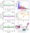
Single-molecule FRET measurements of Bcl-xL refolding. The release of the N-terminal BH4 (α1) helix was inspected at the single-molecule level by fluorescence correlation spectroscopy (FCS) in the presence of 1TOCL:2POPC LUV. Measurements were performed using the same Bcl-xL construct used in Figure 5 between an N-terminally conjugated mCherry fluorescent protein (donor) and acceptor Alexa-Fluor-488 fluorophore introduced at D189C (Figure 2). (ac) Representative snapshots of FCS measurements showed individual fluorescence signals detected for each A488 donor (green) or mCherry acceptor (magenta) fluorophore detected. The presence of a spike appearing simultaneously in both acceptor and donor channels indicated positive FRET events between both fluorophores. The number of FRET events decreased proportionally with the pH of the sample. (d) The single-molecule FRET efficiency (smFRET) in the sample was calculated using Equation (4) from the number of FRET events detected and fitted to a Gaussian distribution. The loss of FRET was characterized by a progressive shift of the distributions to lower FRET efficiencies as a function of pH. This suggested the presence of multiple stable intermediate conformations during the refolding/membrane insertion of Bcl-xL, each with characteristic FRET distances between the BH4 helix and the rest of Bcl-xL. (e) The FRET efficiencies determined by steady-state (Figure 5a), lifetime (Figure 5b), and FCS (Figure 6d) were plotted against experimental pH. (f) Schematic of the experimental set-up, indicating the presence of FRET when the acceptor mCherry was close to the donor A488 and the lack of FRET in the refolded/inserted form of Bcl-xL due to the increase in distance between the donor-acceptor pair. The smFRET data indicated that the release of the BH4 helix was not a two-state transition and involved several Bcl-xL intermediates of various compactness.
Figure 7
Figure 7
Schematic representation of the conformational switching pathway between membrane-anchored and inserted Bcl-xL. After the initial targeting to the MOM via a yet to be fully understood mechanism, Bcl-xL resides in the conformation closely resembling its solution fold, anchored by a single transmembrane helix α8 [16] (top panel). This anchored conformation is distinctly different from the membrane-inserted one (lower panel), characterized by the refolded secondary and tertiary structure, bilayer penetration of various segments (e.g., α6 helix resides about 15 Å from bilayer center [13], and the release of the regulatory BH4 domain (α1 helix) [13]. In this study, we demonstrated that the originally disordered loop between helices α1 and α2 gained helical structure (helix αX) and interacted with the membrane (Figure 3 and Figure 4). Our single-molecule FRET measurements indicated that the insertion transition contained several intermediate states of different compactness (Figure 6). Lipid composition (notably the presence of cardiolipin and other anionic lipids) modulates the propensity of Bcl-xL to undergo protonation-dependent insertion. We hypothesized that conformational switching between the anchored and the inserted conformations of Bcl-xL results in functional switching between canonical and non-canonical (BH4-dependent) modes of apoptotic inhibition (Figure 1b).

Similar articles

Cited by

References

    1. Hardwick J.M., Youle R.J. SnapShot: BCL-2 proteins. Cell. 2009;138:404–404e1. doi: 10.1016/j.cell.2009.07.003. - DOI - PMC - PubMed
    1. Bogner C., Leber B., Andrews D.W. Apoptosis: Embedded in membranes. Curr. Opin. Cell Biol. 2010;22:845–851. doi: 10.1016/j.ceb.2010.08.002. - DOI - PubMed
    1. Kale J., Osterlund E.J., Andrews D.W. BCL-2 family proteins: Changing partners in the dance towards death. Cell Death Differ. 2018;25:65–80. doi: 10.1038/cdd.2017.186. - DOI - PMC - PubMed
    1. Billen L.P., Kokoski C.L., Lovell J.F., Leber B., Andrews D.W. Bcl-XL inhibits membrane permeabilization by competing with Bax. PLoS Biol. 2008;6:e147. doi: 10.1371/journal.pbio.0060147. - DOI - PMC - PubMed
    1. Moldoveanu T., Follis A.V., Kriwacki R.W., Green D.R. Many players in BCL-2 family affairs. Trends Biochem. Sci. 2014;39:101–111. doi: 10.1016/j.tibs.2013.12.006. - DOI - PMC - PubMed

Publication types