Review
. 2020 Feb 26;21(5):1587.
doi: 10.3390/ijms21051587.

Sonic Hedgehog Signaling and Tooth Development

Affiliations
Review

Sonic Hedgehog Signaling and Tooth Development

Akihiro Hosoya et al. Int J Mol Sci. .

Abstract

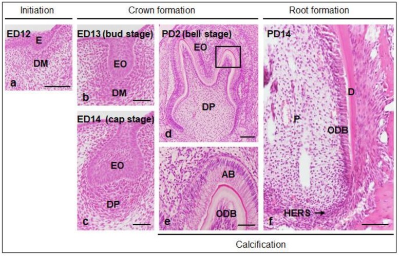
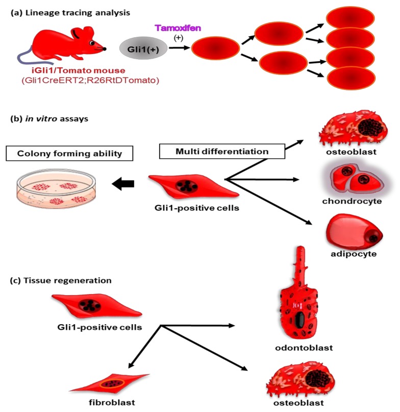
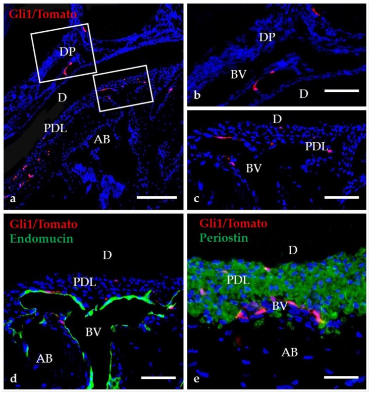
Sonic hedgehog (Shh) is a secreted protein with important roles in mammalian embryogenesis. During tooth development, Shh is primarily expressed in the dental epithelium, from initiation to the root formation stages. A number of studies have analyzed the function of Shh signaling at different stages of tooth development and have revealed that Shh signaling regulates the formation of various tooth components, including enamel, dentin, cementum, and other soft tissues. In addition, dental mesenchymal cells positive for Gli1, a downstream transcription factor of Shh signaling, have been found to have stem cell properties, including multipotency and the ability to self-renew. Indeed, Gli1-positive cells in mature teeth appear to contribute to the regeneration of dental pulp and periodontal tissues. In this review, we provide an overview of recent advances related to the role of Shh signaling in tooth development, as well as the contribution of this pathway to tooth homeostasis and regeneration.

Keywords: Gli1; epithelial and mesenchymal interaction; lineage tracing analysis; mesenchymal stem cell; sonic hedgehog; stem cell marker; tooth development.


Conflict of interest statement

The authors declare no potential conflicts of interest with respect to the authorship and/or publication of this article.

Figures

Figure 1
Figure 1
Process of molar tooth development in mouse. (a–e) Tooth development begins with thickening of the oral epithelium (E) and progresses to crown (bud, cap, and bell stages) and root formation stages. Calcification of enamel, dentin (D), and cementum occurs after the bell stage. The formation stages “initiation,” “crown formation,” and “root formation” correspond to the terms in Table 1. Higher magnification of the boxed region in “d” is shown in “e.” AB, ameloblast; DM, dental mesenchyme; DP, dental papillae; ED, embryonic day; EO, enamel organ; HERS, Hertwig’s epithelial root sheath; ODB, odontoblast; P, pulp; PD, postnatal day. Scale bars = 100 μm (a), 50 μm (bd,f), 25 μm (e).
Figure 2
Figure 2
Schematic illustrations of characteristics of Gli1-positive cells in mature teeth. (a) After tamoxifen administration in iGli1/Tomato mice, Gli1-positive cells are shown to express Tomato red fluorescence. Cells that once expressed Tomato red fluorescence continuously emit this fluorescence even after cell division. Using this system, it is possible to trace the differentiation process of Gli1-positive cells and their progeny cells. (b) Gli1-positive cells exhibit high colony-forming unit fibroblast (CFU-F) activity. These cells also have trilineage potential to form osteoblasts, chondrocytes, and adipocytes in vitro. (c) After tooth transplantation into subcutaneous tissue, Gli1-positive cells differentiate into odontoblasts, osteoblasts, and fibroblasts during tissue regeneration.
Figure 3
Figure 3
Distribution of Gli1-positive cells in mature teeth. Higher magnification of the boxed region in “a” are shown in “b”–“e.” (a–c) Gli1-positive cells are present in the dental pulp (DP) and the periodontal ligament (PDL). (d–e) The merged image of Endomucin and Periostin with Gli1/Tomato fluorescence demonstrate that most Gli1/Tomato-positive cells are distributed near blood vessels (BV). AB, alveolar bone; D, dentin. Scale bars = 100 μm (a), 25 μm (b–e).

References

    1. Kurosaka H. The roles of hedgehog signaling in upper lip formation. Biomed. Res. Int. 2015:901041. doi: 10.1155/2015/901041. - DOI - PMC - PubMed
    1. Xavier G.M., Seppala M., Barrell W., Birjandi A.A., Geoghegan F., Cobourne M.T. Hedgehog receptor function during craniofacial development. Dev. Biol. 2016;415:198–215. doi: 10.1016/j.ydbio.2016.02.009. - DOI - PubMed
    1. Dworkin S., Boglev Y., Owens H., Goldie S.J. The role of Sonic hedgehog in craniofacial patterning, morphogenesis and cranial neural crest survival. J. Dev. Biol. 2016;4:E3. doi: 10.3390/jdb4030024. - DOI - PMC - PubMed
    1. Elliott K.H., Millington G., Brugmann S.A. A novel role for cilia-dependent Sonic hedgehog signaling during submandibular gland development. Dev. Dyn. 2018;247:818–831. doi: 10.1002/dvdy.24627. - DOI - PMC - PubMed
    1. Abramyan J. Hedgehog signaling and embryonic craniofacial disorders. J. Dev. Biol. 2019;7:E2. doi: 10.3390/jdb7020009. - DOI - PMC - PubMed

LinkOut - more resources