. 2020 Feb 26;21(5):1612.
doi: 10.3390/ijms21051612.

ZnO Nanoparticles Induced Caspase-Dependent Apoptosis in Gingival Squamous Cell Carcinoma through Mitochondrial Dysfunction and p70S6K Signaling Pathway

Affiliations

ZnO Nanoparticles Induced Caspase-Dependent Apoptosis in Gingival Squamous Cell Carcinoma through Mitochondrial Dysfunction and p70S6K Signaling Pathway

Shih-Wei Wang L et al. Int J Mol Sci. .

Abstract

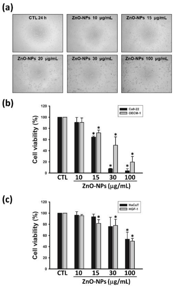
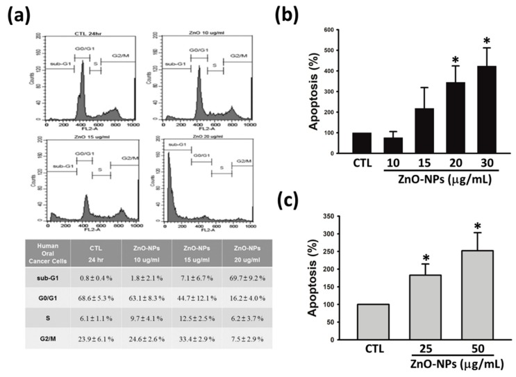
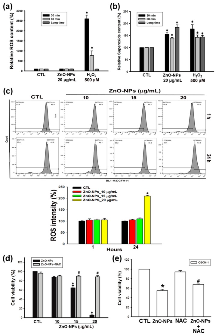
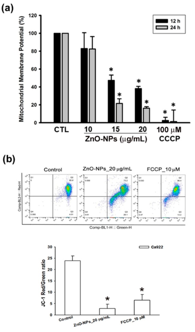
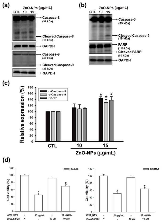
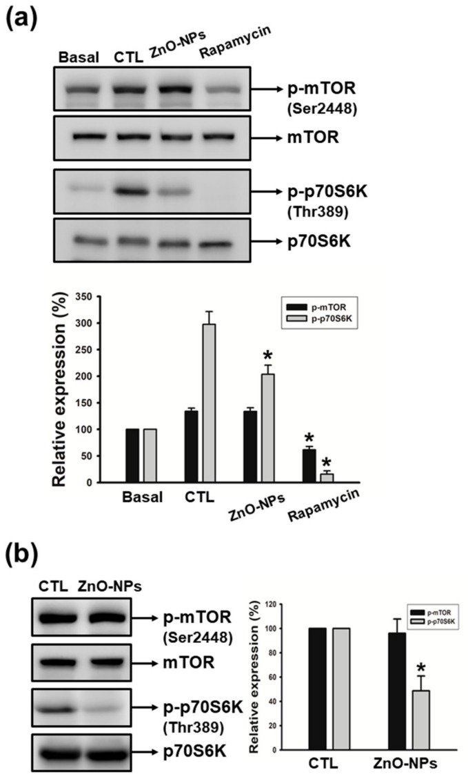
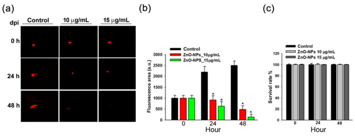
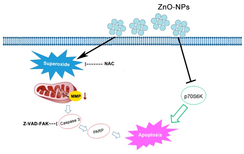
Zinc oxide nanoparticles (ZnO-NPs) are increasingly used in sunscreens, food additives, pigments, rubber manufacture, and electronic materials. Several studies have shown that ZnO-NPs inhibit cell growth and induce apoptosis by the production of oxidative stress in a variety of human cancer cells. However, the anti-cancer property and molecular mechanism of ZnO-NPs in human gingival squamous cell carcinoma (GSCC) are not fully understood. In this study, we found that ZnO-NPs induced growth inhibition of GSCC (Ca9-22 and OECM-1 cells), but no damage in human normal keratinocytes (HaCaT cells) and gingival fibroblasts (HGF-1 cells). ZnO-NPs caused apoptotic cell death of GSCC in a concentration-dependent manner by the quantitative assessment of oligonucleosomal DNA fragmentation. Flow cytometric analysis of cell cycle progression revealed that sub-G1 phase accumulation was dramatically induced by ZnO-NPs. In addition, ZnO-NPs increased the intracellular reactive oxygen species and specifically superoxide levels, and also decreased the mitochondrial membrane potential. ZnO-NPs further activated apoptotic cell death via the caspase cascades. Importantly, anti-oxidant and caspase inhibitor clearly prevented ZnO-NP-induced cell death, indicating the fact that superoxide-induced mitochondrial dysfunction is associated with the ZnO-NP-mediated caspase-dependent apoptosis in human GSCC. Moreover, ZnO-NPs significantly inhibited the phosphorylation of ribosomal protein S6 kinase (p70S6K kinase). In a corollary in vivo study, our results demonstrated that ZnO-NPs possessed an anti-cancer effect in a zebrafish xenograft model. Collectively, these results suggest that ZnO-NPs induce apoptosis through the mitochondrial oxidative damage and p70S6K signaling pathway in human GSCC. The present study may provide an experimental basis for ZnO-NPs to be considered as a promising novel anti‑tumor agent for the treatment of gingival cancer.

Keywords: gingival cancer; p70S6K pathway; superoxide; zinc oxide nanoparticles.


Conflict of interest statement

The authors declare no conflict of interest.

Figures

Figure 1
Figure 1
Effects of zinc oxide nanoparticles (ZnO-NPs) on cell growth in human gingival squamous cell carcinoma (GSCC) and normal cells. Ca9-22 or OECM-1 cells were incubated with the indicated concentrations of ZnO-NPs for 24 h. The cell morphology (a) and viability (b) were assessed by the inverted phase contrast microscope and MTT assay, respectively. (c) Human normal keratinocyte cells (HaCaT) and gingival normal cells (HGF-1) were treated with the indicated concentrations of ZnO-NPs for 24 h, then cell viability was determined using MTT assay. Data represent the mean ± SEM of four independent experiments. * p < 0.05 compared with the control group.
Figure 2
Figure 2
Effects of ZnO-NPs on cell cycle progression and apoptosis in human GSCC cells. (a) Ca9-22 cells were treated with ZnO-NPs (10–20 µg/mL) for 24 h, and subsequently analyzed by propidium iodide (PI) staining to determine the cell cycle distribution. Lower panel, quantitative data were based on histograms. Ca9-22 (b) and OECM-1 (c) cells were treated with the indicated concentrations of ZnO-NPs for 24 h to determine apoptosis using a Cell Death ELISAPLUS kit. Data are expressed as the mean ± SEM of at least three independent experiments. * p < 0.05 compared to the control group.
Figure 3
Figure 3
Effects of ZnO-NPs on the production of reactive oxygen species (ROS) and superoxide in human GSCC cells. (a,b) Ca9-22 cells were incubated with ZnO-NPs for short-term (30–60 min) and long-term (24 h) treatments. Then, the production of ROS and superoxide was examined by ROS/superoxide detection kit. H2O2 was used as a positive control. (c) Ca9-22 cells were treated with ZnO-NPs for the indicated times to determine the intracellular ROS by flow cytometry analysis. Representative flow cytometry-based ROS patterns are shown. The quantification of ROS intensity was analyzed using CellQuest software. (d,e) Ca9-22 and OECM-1 cells were treated with the indicated concentrations of ZnO-NPs with or without N-acetyl-L-cysteine (NAC) (5 mM) for 24 h. Then, the cell viability was determined using MTT assay. Data are expressed as mean ± SEM of four independent experiments. * p < 0.05 compared with control group; # p < 0.05 compared with ZnO-NP-treated group.
Figure 4
Figure 4
Effects of ZnO-NPs on mitochondrial membrane potential of human GSCC cells. (a) Ca9-22 cells were treated with ZnO-NPs (10–20 µg/mL) and carbonyl cyanide m-chlorophenyl hydrazone (CCCP) (100 μM) for the indicated times to determine the change of mitochondrial membrane potential using MITO-ID assay kit. (b) Flow cytometry analysis showed the gating of JC-1 (red) aggregates and JC-1 (green) monomer populations in Ca9-22 cells treated with ZnO-NPs and carbonyl cyanide-4-(trifluoromethoxy)phenylhydrazone (FCCP) for 24 h. Ratio of JC-1 staining represents the mitochondrial function. Data are expressed as mean ± SEM of four independent experiments. * p < 0.05 compared with control group.
Figure 5
Figure 5
Effects of ZnO-NPs on caspase cascade in human GSCC cells. Ca9-22 cells were treated with the indicated concentrations of ZnO-NPs for 24 h. Then, cells were harvested and lysed for the detection of initiator caspase (caspase -8 and -9) (a), caspase-3, and poly-(ADP-ribose) polymerase (PARP) (b) by Western blot analysis. (c) The quantitative densitometry of cleaved form of the indicated caspases and PARP was performed with Image-Pro Plus. (d) Percentage cell viability assessed by MTT assay in GSCC cells, which were treated with ZnO-NPs in the presence or absence of Z-VAD-FAK. Data represent the mean ± SEM of three independent experiments. * p < 0.05 compared with the control group, # p < 0.05 compared to ZnO-NP-treated cells.
Figure 6
Figure 6
Effects of ZnO-NPs on the activation of mTOR and p70S6K in human GSCC cells. (a) Quiescent Ca9-22 cells were treated with or without culture medium (10% fetal bovine serum (FBS)) in the absence (CTL) or presence of ZnO-NPs (20 µg/mL) or rapamycin (10 µM). (b) Quiescent OECM-1 cells were treated with ZnO-NPs (50 µg/mL). Then, cells were harvested and lysed for the detection of p-mTOR and p-p70S6K by Western blot analysis. Image-Pro Plus processing software quantified the relative level of phosphorylated protein. Data represent the mean ± SEM of five independent experiments. * p < 0.05 compared with the control group.
Figure 7
Figure 7
Effects of ZnO-NPs on tumor growth of Ca9-22 cells in zebrafish xenograft model. (a) The intensity of red fluorescence is proportional to the xenograft tumor size. n = 20 embryos for each group. The scale bar is 16× magnification. (b) The quantitative analysis for the anti-tumor efficacy of ZnO-NPs. (c) The survival rate of the zebrafish xenograft model after ZnO-NP treatment is shown. Data represent the mean ± SEM of three independent experiments. * p < 0.05 compared to the vehicle-treated control group.
Figure 8
Figure 8
Schematic diagram of ZnO-NP-induced anti-cancer mechanism in human GSCC. Our study data indicate that ZnO-NPs may promote caspase-dependent apoptosis via mechanisms involving both superoxide-mediated mitochondrial intrinsic and p70S6K signaling pathways.

Similar articles

Cited by

References

    1. Goud E., Malleedi S., Ramanathan A., Wong G.R., Hwei Ern B.T., Yean G.Y., Ann H.H., Syan T.Y., Zain R.M. Association of Interleukin-10 Genotypes and Oral Cancer Susceptibility in Selected Malaysian Population: A Case- Control Study. Asian Pac. J. Cancer Prev. 2019;20:935–941. doi: 10.31557/APJCP.2019.20.3.935. - DOI - PMC - PubMed
    1. Sagheb K., Kumar V., Rahimi-Nedjat R., Dollhausen M., Ziebart T., Al-Nawas B., Walter C. Cervical Metastases Behavior of T1-2 Squamous Cell Carcinoma of the Tongue. J. Maxillofac. Oral. Surg. 2017;16:300–305. doi: 10.1007/s12663-016-0936-0. - DOI - PMC - PubMed
    1. Zhang Y.Y., Wang D.C., Su J.Z., Jia L.F., Peng X., Yu G.Y. Clinicopathological characteristics and outcomes of squamous cell carcinoma of the tongue in different age groups. Head Neck. 2017;39:2276–2282. doi: 10.1002/hed.24898. - DOI - PubMed
    1. Nassiri A.M., Campbell B.R., Mannion K., Sinard R.J., Netterville J.L., Rohde S.L. Survival Outcomes in T4aN0M0 Mandibular Gingival Squamous Cell Carcinoma Treated with Surgery Alone. Otolaryngol. Head Neck Surg. 2019;160:870–875. doi: 10.1177/0194599818821892. - DOI - PubMed
    1. Ramesh R., Sadasivan A. Oral squamous cell carcinoma masquerading as gingival overgrowth. Eur. J. Dent. 2017;11:390–394. doi: 10.4103/ejd.ejd_261_16. - DOI - PMC - PubMed

MeSH terms

Substances

LinkOut - more resources