Comparative Study
. 2020 Feb 28;20(1):42.
doi: 10.1186/s12866-020-1704-3.

The canine oral microbiome: variation in bacterial populations across different niches

Affiliations
Comparative Study

The canine oral microbiome: variation in bacterial populations across different niches

Avika Ruparell et al. BMC Microbiol. .

Abstract

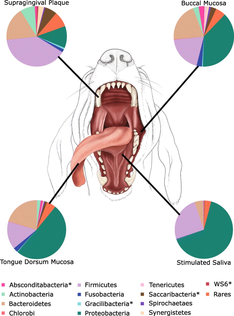
Background: Microbiota from different niches within the canine oral cavity were profiled and compared. Supragingival plaque and stimulated saliva, were collected alongside samples from the buccal and tongue dorsum mucosa, from 14 Labrador retrievers at three timepoints within a 1 month timeframe. The V3-V4 region of the 16S rRNA gene was sequenced via Illumina MiSeq.

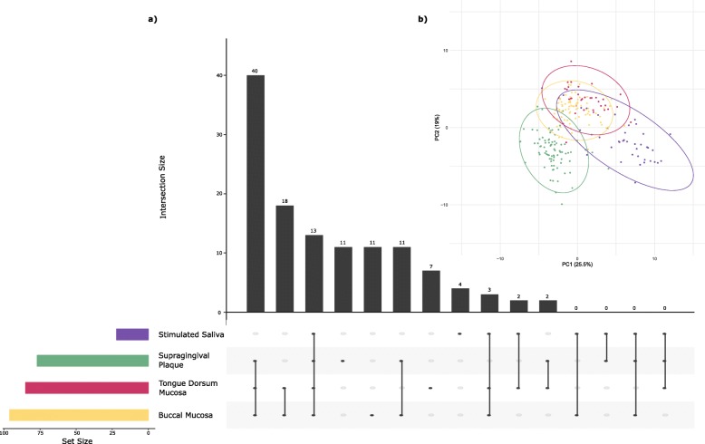
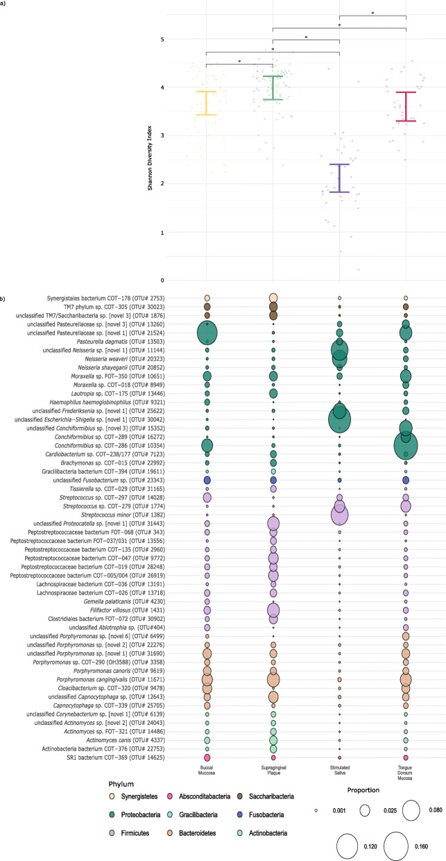
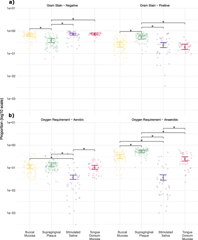
Results: Supragingival plaque microbiota had the highest bacterial diversity and the largest number of significant differences in individual taxa when compared to the other oral niches. Stimulated saliva exhibited the highest variability in microbial composition between dogs, yet the lowest bacterial diversity amongst all the niches. Overall, the bacteria of the buccal and tongue dorsum mucosa were most similar.

Conclusions: The bacterial community profiles indicated three discrete oral niches: soft tissue surfaces (buccal and tongue dorsum mucosa), hard tissue surface (supragingival plaque) and saliva. The ability to distinguish the niches by their microbiota signature offers the potential for microbial biomarkers to be identified in each unique niche for diagnostic use.

Keywords: Buccal; Canine; Microbiome; Oral; Plaque; Saliva; Tongue.


Conflict of interest statement

All authors were employees of WALTHAM Petcare Science Institute (Mars Petcare) at the time of the study and declare that they have no competing interests.

Figures

Fig. 1
Fig. 1
(a) UpSet plot based on presence/absence of operational taxonomic units where presence > 0.5% average OTU abundance mapped alongside (b) principal component scores with 95% confidence regions from analysis performed on the log10 proportions of operational taxonomic units identified in each of the oral niches: Buccal mucosa (yellow), supragingival plaque (green), saliva (purple) and tongue dorsum mucosa (magenta)
Fig. 2
Fig. 2
Average phylogenetic distribution of operational taxonomic units based on sequence reads across the canine oral niches. Asterisks indicate candidate phyla. Central image: © Emily McDougall Art
Fig. 3
Fig. 3
(a) Shannon diversity index with 95% confidence intervals (b) plotted against bacterial species indicating significant differences between the four niches within the dogs’ mouth. (b) indicates operational taxomomic units with abundance > 0.5% for at least one of the oral niches, where the size of the circles represent the proportion and the colours represent the phylum
Fig. 4
Fig. 4
Proportions of bacterial species per sample discriminated by (a) Gram-stain status and (b) oxygen requirements. Coloured bars indicate mean proportions of OTUs with 95% confidence intervals across the canine oral niches: Buccal mucosa (yellow), supragingival plaque (green), saliva (purple) and tongue dorsum mucosa (magenta). Black bars and asterisks indicate niches with significant differences (all p < 0.001)

Similar articles

Cited by

References

    1. Dewhirst FE, Klein EA, Thompson EC, Blanton JM, Chen T, Milella L, Buckley CM, Davis IJ, Bennett ML, Marshall-Jones ZV. The canine oral microbiome. PLoS One. 2012;7(4):e36067. doi: 10.1371/journal.pone.0036067. - DOI - PMC - PubMed
    1. Dewhirst FE, Chen T, Izard J, Paster BJ, Tanner AC, Yu WH, Lakshmanan A, Wade WG. The human oral microbiome. J Bacteriol. 2010;192(19):5002–5017. doi: 10.1128/JB.00542-10. - DOI - PMC - PubMed
    1. Sorensen WP, Loe H, Ramfjord SP. Periodontal disease in the beagle dog. A cross sectional clinical study. J Periodontal Res. 1980;15(4):380–389. doi: 10.1111/j.1600-0765.1980.tb00295.x. - DOI - PubMed
    1. Lindhe J, Hamp S, Loe H. Experimental periodontitis in the beagle dog. J Periodontal Res. 1973;8(1):1–10. doi: 10.1111/j.1600-0765.1973.tb00735.x. - DOI - PubMed
    1. Harvey CE. Periodontal disease in dogs. Etiopathogenesis, prevalence, and significance. Vet Clin North Am Small Anim Pract. 1998;28(5):1111–1128. doi: 10.1016/S0195-5616(98)50105-2. - DOI - PubMed

Publication types