. 2020 Feb 28;11(1):91.
doi: 10.1186/s13287-020-01612-y.

Mesenchymal stem cells regulate the Th17/Treg cell balance partly through hepatocyte growth factor in vitro

Affiliations

Mesenchymal stem cells regulate the Th17/Treg cell balance partly through hepatocyte growth factor in vitro

Qi-Hong Chen et al. Stem Cell Res Ther. .

Abstract

Introduction: Mesenchymal stem cells (MSCs) exert immunomodulatory functions by inducing the development and differentiation of naive T cells into T cells with an anti-inflammatory regulatory T cell (Treg) phenotype. Our previous study showed that hepatocyte growth factor (HGF) secreted by MSCs had immunomodulatory effects in the context of lipopolysaccharide (LPS) stimulation. We hypothesized that HGF is a key factor in the MSC-mediated regulation of the T helper 17 (Th17) cell/regulatory T (Treg) cell balance.

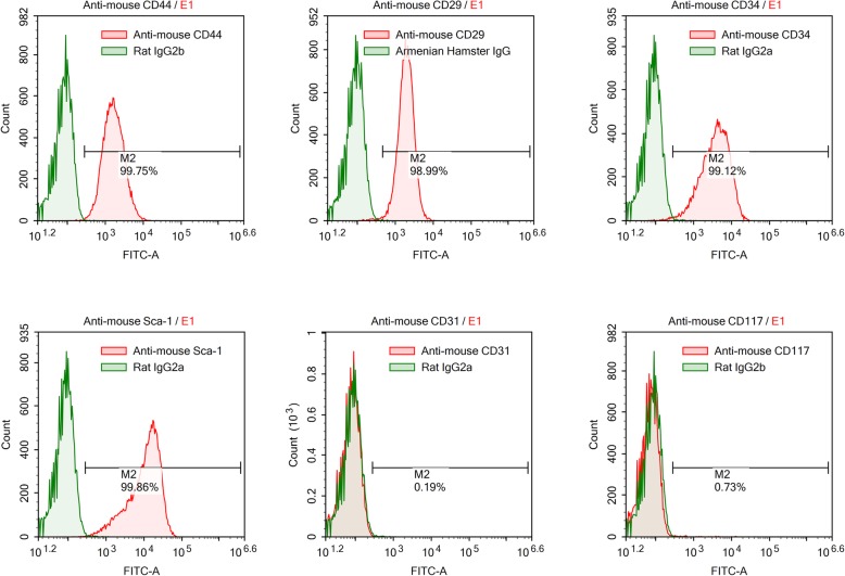
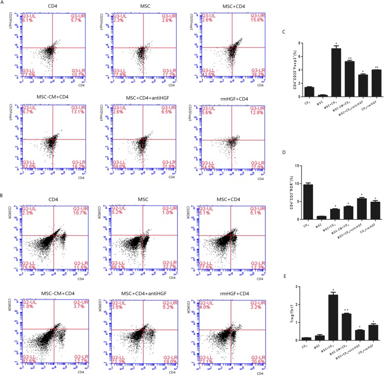
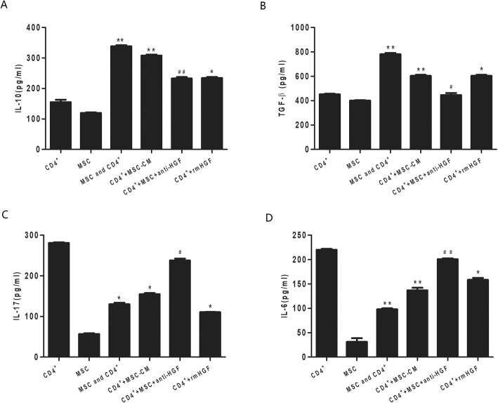
Methods: We investigated the effects of MSCs on the differentiation of CD4+ T cells and the functions of Th17/Treg cells in response to LPS stimulation by performing in vitro coculture experiments. MSCs were added to the upper chambers of cell culture inserts, and CD4+ T cells were plated in the lower chambers, followed by treatment with LPS or an anti-HGF antibody. Th17 (CD4+CD3+RORrt+) and Treg (CD4+CD25+Foxp3+) cell frequencies were analysed by flow cytometry, and the expression of Th17 cell- and Treg cell-related cytokines in the CD4+ T cells or culture medium was measured by quantitative PCR (qPCR) and enzyme-linked immunosorbent assay (ELISA), respectively. Neutrophil functions were determined by flow cytometry after a coculture with Th17/Treg cells.

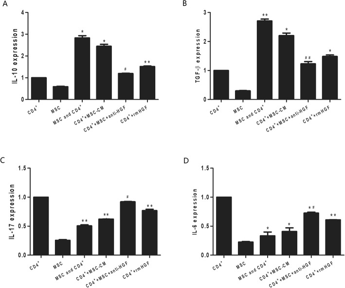
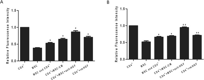
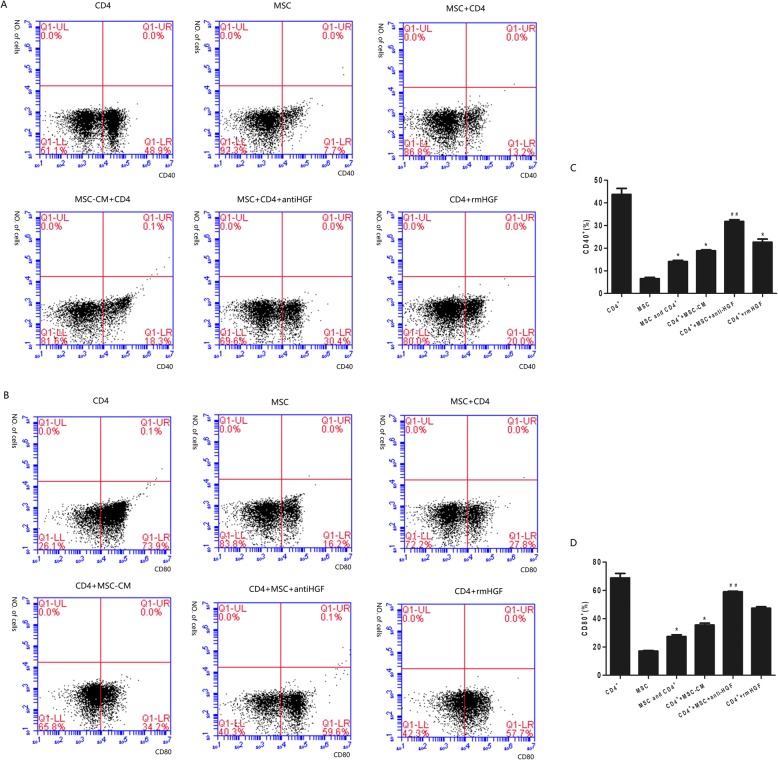
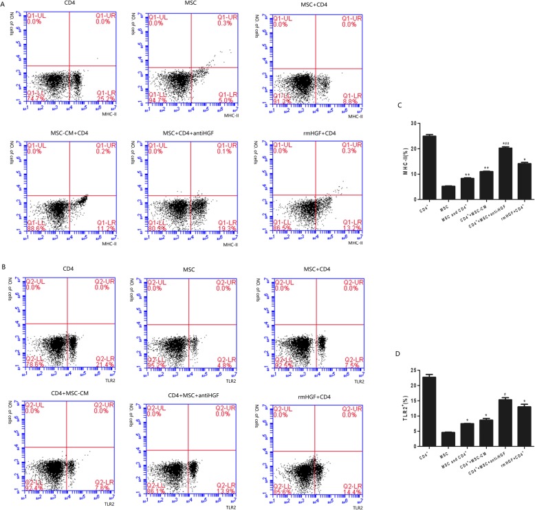
Results: The percentage of CD4+CD25+Foxp3+ cells was significantly increased in the CD4+ T cell population, while the percentage of CD4+CD3+RORrt+ cells was significantly decreased after MSC coculture. However, the MSC-induced effect was significantly inhibited by the anti-HGF antibody (p < 0.05). Furthermore, MSCs significantly inhibited the CD4+ T cell expression of IL-17 and IL-6 but increased the expression of IL-10 (p < 0.05 or p < 0.01); these effects were inhibited by the anti-HGF antibody (p < 0.05). In addition, CD4+ T cells cocultured with MSCs significantly inhibited neutrophil phagocytic and oxidative burst activities (p < 0.05 or p < 0.01); however, these MSC-induced effects were inhibited by the anti-HGF antibody (p < 0.05).

Conclusion: These data suggested that MSCs induced the conversion of fully differentiated Th17 cells into functional Treg cells and thereby modulated the Th17/Treg cell balance in the CD4+ T cell population, which was partly attributed to HGF secreted by the MSCs.

Keywords: Hepatocyte growth factor; Mesenchymal stem cells; Th17; Treg.


Conflict of interest statement

The authors declare that they have no competing interests.

Figures

Fig. 1
Fig. 1
Flow cytometry identification of mouse mesenchymal stem cells (MSCs). Cell surface markers of MSCs, including CD44, CD29, CD117, CD31, CD34 and Sca-1, were analysed with flow cytometry. Red lines represent the isotype controls. Cytofluorometric analyses showed the presence of several molecules such as CD44, CD29, CD34 and Sca-1 but not the presence of CD117 or CD31
Fig. 2
Fig. 2
The modulatory effect of HGF secreted by MSCs on Th17/Treg cell differentiation in CD4+ T cells analysed by flow cytometry. The results showed that the percentage of CD4+CD25+Foxp3+ cells was significantly increased in the CD4+ T cell population, but the MSC-induced effect was significantly inhibited by an anti-HGF antibody (p < 0.05, a, c). The percentage of CD4+CD3+RORrt+ cells was significantly decreased after MSC coculture. However, the MSC-induced effect was significantly inhibited by the anti-HGF antibody (p < 0.05, b, d) (n = 3; *p < 0.05 or **p < 0.01 vs. the CD4+ group; #p < 0.05 vs. the MSC+CD4+ group). MSCs, mesenchymal stem cells; rmHGF, recombinant mouse hepatocyte growth factor
Fig. 3
Fig. 3
The regulatory effect of HGF secreted by MSCs on cytokine mRNA expression in CD4+ T cells analysed by qPCR. The results showed that MSCs significantly increased IL-10 (p < 0.05, a) and TGF-β (p < 0.01, b) mRNA expression and inhibited IL-17 (p < 0.01, c) and IL-6 (p < 0.05, d) mRNA expression in CD4+ T cells. However, the MSC-induced effects were inhibited by an anti-HGF antibody (p < 0.05 or p < 0.01) (n = 3; *p < 0.05 or **p < 0.01 vs. the CD4+ group; #p < 0.05 or ##p < 0.01 vs. the MSC+CD4+ group). MSCs, mesenchymal stem cells; rmHGF, recombinant mouse hepatocyte growth factor; qPCR, quantitative polymerase chain reaction
Fig. 4
Fig. 4
The regulatory effect of HGF secreted by MSCs on cytokine production in CD4+ T cells detected by ELISA. We found that MSCs significantly increased the CD4+ T cell secretion of IL-10 (p < 0.01, a) and TGF-β (p < 0.01, b) but inhibited the CD4+ T cell secretion of IL-17 (p < 0.05, c) and IL-6 (p < 0.01, d). However, the MSC-induced effects were inhibited by an anti-HGF antibody (p < 0.05 or p < 0.01) (n = 3; *p < 0.05 or **p < 0.01 vs. the CD4+ group; #p < 0.05 or ##p < 0.01 vs. the MSC+CD4+ group). MSCs, mesenchymal stem cells; rmHGF, recombinant mouse hepatocyte growth factor
Fig. 5
Fig. 5
The regulatory effect of HGF secreted by MSCs on neutrophil activities in cells cocultured with CD4+ T cells and analysed with flow cytometry. The results indicated that CD4+ T cells cocultured with MSCs significantly inhibited neutrophil phagocytic (p < 0.05, a) and oxidative burst activities (p < 0.01, b). However, the effects of the CD4+ T cells were inhibited by an anti-HGF antibody (p < 0.05 or p < 0.01) (n = 3; *p < 0.05 or **p < 0.01 vs. the CD4+ group; #p < 0.05 or ##p < 0.01 vs. the MSC+CD4+ group). MSCs, mesenchymal stem cells; rmHGF, recombinant mouse hepatocyte growth factor
Fig. 6
Fig. 6
The regulatory effect of HGF secreted by MSCs on CD40+ and CD80+ monocyte activation in cocultures with CD4+ T cells and analysed with flow cytometry. The results showed that after being cocultured with MSCs, CD4+ T cells significantly inhibited monocyte expression of CD40 (p < 0.05, a) and CD80 (p < 0.05, b). However, the CD4+ T cell-mediated effect was inhibited by an anti-HGF antibody (p < 0.01) (n = 3; *p < 0.05 vs. the CD4+ group; ##p < 0.01 vs. the MSC+CD4+ group). MSCs, mesenchymal stem cells; rmHGF, recombinant mouse hepatocyte growth factor
Fig. 7
Fig. 7
The regulatory effect of HGF secreted by MSCs on MHC-II and TLR-2 expression in monocytes cocultured with CD4+ T cells and analysed with flow cytometry. The results showed that after being cocultured with MSCs, CD4+ T cells significantly inhibited monocyte expression of MHC-II (p < 0.01, a) and TLR2 (p < 0.05, b). However, the CD4+ T cell-mediated effect was inhibited by an anti-HGF antibody (p < 0.05 or p < 0.01) (n = 3; *p < 0.05 or **p < 0.01 vs. the CD4+ group; ##p < 0.01 vs. the MSC+CD4+ group). MSCs, mesenchymal stem cells; rmHGF, recombinant mouse hepatocyte growth factor

References

    1. Villar J, Zhang H, Slutsky AS. Lung repair and regeneration in ARDS: role of PECAM1 and Wnt signaling. Chest. 2019;155(3):587–594. doi: 10.1016/j.chest.2018.10.022. - DOI - PMC - PubMed
    1. Matthay MA, Zemans RL, Zimmerman GA, et al. Acute respiratory distress syndrome. Nat Rev Dis Primers. 2019;5(1):18. doi: 10.1038/s41572-019-0069-0. - DOI - PMC - PubMed
    1. Reiss LK, Schuppert A, Uhlig S. Inflammatory processes during acute respiratory distress syndrome: a complex system. Curr Opin Crit Care. 2018;24(1):1–9. doi: 10.1097/MCC.0000000000000472. - DOI - PubMed
    1. Adamzik M, Broll J, Steinmann J, Westendorf AM, et al. An increased alveolar CD4 + CD25 + Foxp3 + T-regulatory cell ratio in acute respiratory distress syndrome is associated with increased 30-day mortality. Intensive Care Med. 2013;39(10):1743–1751. doi: 10.1007/s00134-013-3036-3. - DOI - PMC - PubMed
    1. Zhu G, Liu Y, Zhang W, et al. CD27+TIM-1+ memory B cells promoted the development of Foxp3+ Tregs and were associated with better survival in acute respiratory distress syndrome. Immunol Res. 2018;66(2):281–287. doi: 10.1007/s12026-017-8983-2. - DOI - PubMed

Publication types

Substances