. 2020 Apr 17;86(9):e02950-19.
doi: 10.1128/AEM.02950-19. Print 2020 Apr 17.

Biofilm Interactions of Candida albicans and Mitis Group Streptococci in a Titanium-Mucosal Interface Model

Affiliations

Biofilm Interactions of Candida albicans and Mitis Group Streptococci in a Titanium-Mucosal Interface Model

João Gabriel Silva Souza et al. Appl Environ Microbiol. .

Abstract

Streptococci from the mitis group (represented mainly by Streptococcus mitis, Streptococcus oralis, Streptococcus sanguinis, and Streptococcus gordonii) form robust biofilms with Candida albicans in different experimental models. These microorganisms have been found in polymicrobial biofilms forming on titanium biomaterial surfaces in humans with peri-implant disease. The purpose of this work was to study mutualistic interactions in biofilms forming on titanium and their effect on the adjacent mucosa, using a relevant infection model. Single and mixed biofilms of C. albicans and each Streptococcus species were grown on titanium disks. Bacterial and fungal biovolume and biomass were quantified in these biofilms. Organotypic mucosal constructs were exposed to preformed titanium surface biofilms to test their effect on secretion of proinflammatory cytokines and cell damage. C. albicans promoted bacterial biofilms of all mitis Streptococcus species on titanium surfaces. This relationship was mutualistic since all bacterial species upregulated the efg1 hypha-associated gene in C. albicans Mixed biofilms caused increased tissue damage but did not increase proinflammatory cytokine responses compared to biofilms comprising Candida alone. Interestingly, spent culture medium from tissues exposed to titanium biofilms suppressed Candida growth on titanium surfaces.IMPORTANCE Our findings provide new insights into the cross-kingdom interaction between C. albicans and Streptococcus species representative of the mitis group. These microorganisms colonize titanium-based dental implant materials, but little is known about their ability to cause inflammation and damage of the adjacent mucosal tissues. Using an in vitro biomaterial-mucosal interface infection model, we showed that mixed biofilms of each species with C. albicans enhance tissue damage. One possible mechanism for this effect is the increased fungal hypha-associated virulence gene expression we observed in mixed biofilms with these species. Interestingly, we also found that the interaction of multispecies biofilms with organotypic mucosal surfaces led to the release of growth-suppressing mediators of Candida, which may represent a homeostatic defense mechanism of the oral mucosa against fungal overgrowth. Thus, our findings provide novel insights into biofilms on biomaterials that may play an important role in the pathogenesis of mucosal infections around titanium implants.

Keywords: Candida albicans; Streptococcus; biofilms; peri-implant mucositis; peri-implantitis.


Figures

FIG 1
FIG 1
Single-species biofilms growing for 72 h on titanium surfaces. (A) x-y isosurfaces (top) and three-dimensional reconstructions (bottom) of representative confocal laser scanning microscopy images of biofilms. Bacteria (red) were visualized after fluorescence in situ hybridization with an all-bacterial (EUB) probe labeled with Alexa 633. Candida albicans (green) was visualized after staining with an FITC-conjugated anti-Candida antibody. Scale bars, 50 μm (x-y isosurfaces) and 70 μm (three-dimensional reconstructions). (B) Average total biovolumes (in μm3). Biovolumes were measured in two different confocal laser scanning microscopy image stacks from two independent experiments. For one-way ANOVA, different letters represent statistical differences among the groups (P < 0.05). (C) Average microorganism count in logarithmic scale using qPCR of 16S or 18S rRNA genes. The error bars indicate standard deviations. Ca, C. albicans; So, S. oralis; Sg, S. gordonii; Sm, S. mitis; Ss, S. sanguinis.
FIG 2
FIG 2
Seventy-two-hour mixed biofilms of C. albicans with Streptococcus species on titanium surfaces. (A) x-y isosurfaces (top) and three-dimensional reconstructions (bottom) of representative confocal laser scanning microscopy images of biofilms. Bacteria (red) were visualized after fluorescence in situ hybridization with an all-bacterial (EUB) probe labeled with Alexa 633. Candida albicans (green) was visualized after staining with an FITC-conjugated anti-Candida antibody. Scale bars, 50 μm (x-y isosurfaces) and 70 μm (three-dimensional reconstructions). (B) Average total biovolumes (in μm3) for each mixed biofilm. Biovolumes were measured in two different confocal laser scanning microscopy image stacks from two independent experiments. (C) Bacterial biomass (qPCR of 16S rRNA gene) expressed as fold of mixed over single biofilms. (D) Average Candida biovolumes in single and mixed biofilms (in μm3). For one-way ANOVA, different letters represent statistical differences among the groups (P < 0.05). (E) Candida biomass (qPCR of 18S rRNA gene) expressed as fold of mixed over single biofilms. (F) Relative expression of Candida genes assessed by RT-qPCR. Results represent the mean fold change in gene expression in mixed biofilms over Candida alone in three independent experiments. *, P < 0.05 using the Bonferroni t test. The error bars indicate standard deviations. Ca, C. albicans; So, S. oralis; Sg, S. gordonii; Sm, S. mitis; Ss, S. sanguinis.
FIG 3
FIG 3
Twenty-four-hour Candida biofilm growth on preformed Streptococcus biofilms on titanium surfaces. Streptococcal biofilms were grown for 48 h prior to C. albicans inoculation. (A) x-y isosurfaces (top) and three-dimensional reconstructions (bottom) of representative confocal laser scanning microscopy images of biofilms. Bacteria (red) and Candida albicans (green) were visualized by staining as described for Fig. 1 and 2. (B) Average of total, Candida, and bacterial biovolumes in single and mixed biofilms. (C) Candida biomass by qPCR. For one-way ANOVA, different letters represent statistical differences among the groups (P < 0.05). (D) Bacterial biomass by qPCR. *, P < 0.05 using the Bonferroni t test. The error bars indicate standard deviations. Ca, C. albicans; So, S. oralis; Sg, S. gordonii; Sm, S. mitis; Ss, S. sanguinis.
FIG 4
FIG 4
Preformed (72-h) titanium biofilms of C. albicans alone or in combination with Streptococcus species, juxtaposed to organotypic mucosal surfaces for 16 h. (A) Representative tissue sections (top) of mucosal surfaces visualized after staining for bacteria (red) with an all-bacterial (EUB) probe labeled with Alexa 633. Notice the absence of bacteria on the mucosal surface. Candida albicans (green) was visualized after staining with an FITC-conjugated anti-Candida antibody, and mucosal cell nuclei were counterstained with the nucleic acid stain Hoechst 33258 (blue). Corresponding hematoxylin- and eosin-stained tissue sections are shown in the bottom panels. (B) Lactate dehydrogenase released by mucosal analogs after 16-h exposure to preformed titanium biofilms. Abs, absorbance. (C) Cytokines released from organotypic mucosa tissues after 16-h exposure to preformed titanium biofilms. Interleukin-1β (IL-1β), interleukin-6 (IL-6), interleukin-8 (IL-8), and tumor necrosis factor alpha (TNFα) concentrations were simultaneously quantified by multiplex enzyme-linked immunosorbent assay (ELISA). For one-way ANOVA, different letters represent statistical differences among the groups (P < 0.05). The error bars indicate standard deviations. Ca, C. albicans; So, S. oralis; Sg, S. gordonii; Sm, S. mitis; Ss, S. sanguinis.
FIG 5
FIG 5
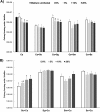
Effect of mucosal tissue spent media on C. albicans growth on titanium surfaces. Biofilm media were supplemented with increasing concentrations (0, 5, 10, and 20%) of spent media collected from organotypic mucosae after exposure to respective preformed 72-h titanium biofilms. Spent media from organotypic mucosal tissues exposed to sterile titanium disks (black solid bar) were used as a control. (A) Candida viable counts expressed as log10 CFU. (B) Bacterial viable counts expressed as log10 CFU. For one-way ANOVA, different letters represent statistical differences among the groups (P < 0.05). The error bars indicate standard deviations. Ca, C. albicans; So, S. oralis; Sg, S. gordonii; Sm, S. mitis; Ss, S. sanguinis.

References

    1. Costerton JW, Lewandowski Z, Caldwell DE, Korber DR, Lappin-Scott HM. 1995. Microbial biofilms. Annu Rev Microbiol 49:711–745. doi:10.1146/annurev.mi.49.100195.003431. - DOI - PubMed
    1. Marsh PD, Moter A, Devine DA. 2011. Dental plaque biofilms: communities, conflict and control. Periodontol 2000 55:16–35. doi:10.1111/j.1600-0757.2009.00339.x. - DOI - PubMed
    1. Lang NP, Brägger U, Walther D, Beamer B, Kornman KS. 1993. Ligature-induced peri-implant infection in cynomolgus monkeys. I. Clinical and radiographic findings. Clin Oral Implants Res 4:2–11. doi:10.1034/j.1600-0501.1993.040101.x. - DOI - PubMed
    1. Pontoriero R, Tonelli MP, Carnevale G, Mombelli A, Nyman SR, Lang NP. 1994. Experimentally induced peri-implant mucositis. A clinical study in humans. Clin Oral Implants Res 5:254–259. doi:10.1034/j.1600-0501.1994.050409.x. - DOI - PubMed
    1. Salvi GE, Cosgarea R, Sculean A. 2017. Prevalence and mechanisms of peri-implant diseases. J Dent Res 96:31–37. doi:10.1177/0022034516667484. - DOI - PubMed

Publication types

LinkOut - more resources