. 2020 Nov;69(11):1928-1938.
doi: 10.1136/gutjnl-2019-319523. Epub 2020 Feb 28.

Mitochondrial dysfunction during loss of prohibitin 1 triggers Paneth cell defects and ileitis

Affiliations

Mitochondrial dysfunction during loss of prohibitin 1 triggers Paneth cell defects and ileitis

Dakota N Jackson et al. Gut. 2020 Nov.

Abstract

Objective: Although perturbations in mitochondrial function and structure have been described in the intestinal epithelium of Crohn's disease and ulcerative colitis patients, the role of epithelial mitochondrial stress in the pathophysiology of inflammatory bowel diseases (IBD) is not well elucidated. Prohibitin 1 (PHB1), a major component protein of the inner mitochondrial membrane crucial for optimal respiratory chain assembly and function, is decreased during IBD.

Design: Male and female mice with inducible intestinal epithelial cell deletion of Phb1 (Phb1iΔIEC ) or Paneth cell-specific deletion of Phb1 (Phb1ΔPC ) and Phb1fl/fl control mice were housed up to 20 weeks to characterise the impact of PHB1 deletion on intestinal homeostasis. To suppress mitochondrial reactive oxygen species, a mitochondrial-targeted antioxidant, Mito-Tempo, was administered. To examine epithelial cell-intrinsic responses, intestinal enteroids were generated from crypts of Phb1iΔIEC or Phb1ΔPC mice.

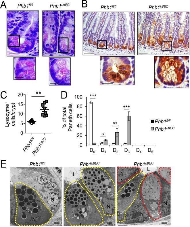
Results: Phb1iΔIEC mice exhibited spontaneous ileal inflammation that was preceded by mitochondrial dysfunction in all IECs and early abnormalities in Paneth cells. Mito-Tempo ameliorated mitochondrial dysfunction, Paneth cell abnormalities and ileitis in Phb1iΔIEC ileum. Deletion of Phb1 specifically in Paneth cells (Phb1ΔPC ) was sufficient to cause ileitis. Intestinal enteroids generated from crypts of Phb1iΔIEC or Phb1ΔPC mice exhibited decreased viability and Paneth cell defects that were improved by Mito-Tempo.

Conclusion: Our results identify Paneth cells as highly susceptible to mitochondrial dysfunction and central to the pathogenesis of ileitis, with translational implications for the subset of Crohn's disease patients exhibiting Paneth cell defects.

Keywords: crohn's disease; epithelial cells; inflammatory bowel disease.


Conflict of interest statement

Competing interests: None declared.

Figures

Figure 1
Figure 1
Phb1i ΔIEC mice develop spontaneous ileitis 12 weeks after Phb1 deletion. (A) H&E-stained sections showing ileum histology. Scale bars: 250 µm, boxed pullout: 75 µm. (B) The number of epithelial cells across 50 crypts or 50 villi per mouse. (C) Absolute number of ileal lamina propria (LP) immune cells calculated of total LP cells isolated from ileum. Data are representative of three independent experiments. (D) Representative flow cytometry plots gated on CD45+ cells (top) or gated on CD45+MHCII+Gr-1-B220-Cells (bottom) (E) mRNA expression in ileum measured by qPCR. Results are presented as individual data points±SEM of 10 mice (B), 6-7 mice (1 Phb1i ΔIEC outlier removed after Grubbs’ test) (C), or 6 mice (E) per group. *P<0.05, **P<0.01, ***P<0.001 by unpaired, two-tailed Student’s t-test. IFN-γ, interferon-γ; IL-10, interleukin-10; Phb1i ΔIEC, intestinal epithelial cell deletion of Phb1; Phb1 ΔPC, Paneth cell-specific deletion of Phb1; TNFα, tumour necrosis factor α.
Figure 2
Figure 2
Phb1iΔIEC ileal IECs exhibit mitochondrial dysfunction. Time point shown is 12 weeks after Phb1 deletion. (A) TEM of ileal enterocyte. Representative healthy (blue box), unhealthy (orange box), and dense inclusion body-containing (red box) mitochondria. Scale bars: 500 nm. N, nucleus. (B) % healthy, unhealthy, and dense inclusion body-containing mitochondria visualised by TEM. n=200 enterocytes, 200 goblet cells, 50 Paneth cells, and 50 CBC cells with an average of 42 mitochondrial/cell. Enteroendocrine cells were too sparse to quantitate. (C) ETC complex activity in isolated ileal IECs. (D) Representative Western blots of mtUPR markers in isolated ileal IECs. Results are presented as pooled data means±SEM (B) or as individual data points±SEM (C) of four mice per group (B) of eight mice per group (C, D). *P<0.05, **P<0.01 by 1-way ANOVA followed by Bonferroni’s test (B) or by unpaired, 2-tailed Student’s t-test (C). ANOVA, analysis of variance; CBC, crypt base columnar; ETC, electron transport chain; HSP, heat shock protein; IECs, intestinal epithelial cells; mtUPR, mitochondrial unfolded protein response; Phb1iΔIEC, intestinal epithelial cell deletion of Phb1; PKR, protein kinase R; SEM, standard error mean; TEM, transmission electron microscopy.
Figure 3
Figure 3
Phb1ΔIEC mice exhibit ileal Paneth cell abnormalities. Time point shown is 12 weeks afetr Phb1 deletion. (A) H&E-stained sections showing Paneth cells. Scale bars: 50 µm, boxed pullout: 20 µm. (B) Lysozyme IHC staining. Scale bars: 250 µm, boxed pullouts: 50 µm (C) The number of lysozyme+ cells across 50 crypts per mouse. (D) % of Paneth cells displaying each pattern of lysozyme expression. D0, normal; D1, disordered; D2, diminished; D3, diffuse; D4, excluded; D5, enlarged. 500 cells quantitated each mouse. (E) TEM of crypt base. Paneth cells, yellow outline; goblet-like cells, red outline. N, nucleus; L, lumen. Scale bars: 2 µm. Results are presented as individual data points±SEM (C) or as pooled data means± SEM (D) of 8 mice per group. **P<0.01, ***p<0.001 by unpaired, 2-tailed Student’s t-test. (B) Or by 1-way ANOVA followed by Bonferroni’s test (D). ANOVA, analysis of variance; Phb1 ΔIEC, intestinal epithelial cell deletion of Phb1.
Figure 4
Figure 4
Ileal Paneth cells resemble goblet/Paneth intermediate cells and goblet cell number and size are increased in Phb1 iΔIEC mice. Time point shown is 12 weeks after Phb1 deletion (A) AB-PAS-stained sections. Dashed line denotes crypt base. Scale bars: 250 μm, boxed pullouts: 50 μm. (B) The number of AB+ cells in the crypt base (below dashed line in A) or above the crypt base across 50 crypts per mouse. (C) MUC2 (goblet cell marker) and Lysozyme (Paneth cell marker) immunofluorescene staining. Scale bars: 250 μm, boxed pullout showing co-localisation: 75 μm. (D) Quantitation of average mucin area/goblet cell above the crypt base. 250 cells quantitated each mouse. (E) TEM of villus goblet cells. Mucin granules, yellow outline. Scale bars: 1 μm. Results are presented as individual data points ± SEM of 8 mice per group. **P<0.01, ***p<0.001 by unpaired, 2-tailed student’s t test. AB-PAS, alcian blue–periodic acid schiff; L, lumen; MUC2, mucin2; N, nucleus; Phb1 iΔIEC, intestinal epithelial cell deletion of Phb1; SEM, standard error mean; TEM, transmission electron microscopy.
Figure 5
Figure 5
Mito-Tempo (MT) ameliorates mitochondrial dysfunction, Paneth cells defects, and ileitis in Phb1 iΔIEC ileum. (A) mRNA expression of mtUPR (ClpP, Hsp60) and ER UPR (sXbp1, BiP, Grp94) markers in isolated ileal IECs measured by qPCR. (B) TEM of ileal enterocytes. Arrows indicate mitochondria. Scale bars: 500 nm. (C) % healthy, unhealthy (swollen, dissolution of cristae), and dense inclusion body-containing mitochondria visualized by TEM. n=200 enterocytes, 200 goblet cells, 50 Paneth cells, and 50 CBC cells with an average of 53 mitochondrial/cell. (D) AB-PAS and Lysozyme immunohistochemistry staining. Scale bars: 250 μm. mRNA expression in isolated ileal IECs (E) or whole ileum (F) measured by qPCR. (G) H&E-stained sections showing ileum histology at 12 weeks. Scale bars: 250 μm. Results are presented as pooled data means±SEM of 6 mice per group (A, E, F) or 4 mice per group (C). *P<0.05, **p<0.01 vs Phb1 fl/fl veh, #p<0.05 vs Phb1 iΔIEC veh by 1-way ANOVA followed by Bonferroni’s test. AB-PAS, alcian blue–periodic acid schiff; ANOVA, analysis of variance; CBC, crypt base columnar; ER, endoplasmic reticulum; IECs, intestinal epithelial cells; IFN-γ, interferon-γ; IL-18, interleukin-18; mtUPR, mitochondrial unfolded protein response; Phb1 iΔIEC, intestinal epithelial cell deletion of Phb1; TEM, transmission electron microscopy; TNFα, tumour necrosis factor α.
Figure 6
Figure 6
Phb1 Defα6ΔC mice develop mitochondrial dysfunction, Paneth cell defects, and spontaneous ileitis. (A) H&E-stained ileum sections at 20 weeks of age. Scale bars: 250 μm. (B) Absolute number of ileal LP immune cells calculated of total ileal LP cells. (C) mRNA expression in ileum measured by qPCR. (D) The number of Lysozyme+ cells across 50 crypts per mouse. (E) % of Paneth cells displaying each pattern of Lysozyme expression. 50 cells quantitated each mouse. (F) The number of AB+ cells in the crypt base or above the crypt base across 50 crypts per mouse. (G) TEM of Phb1 Defα6ΔPC ileum crypt base. Paneth cells, yellow outline; goblet-like cells, red outline; Paneth cell with vesiculated ER, green outline. Scale bars: 1 μm. Box denotes area of higher magnification showing unhealthy mitochondria. (H) % of Paneth cells displaying vesiculated ER. Results are presented as individual data points ± SEM (B, D, F) or as pooled data means ± SEM (C, E) of 9 mice per group. *P<0.05, **p<0.01, ***p<0.001 vs PHB1fl/fl by unpaired, 2-tailed Student’s t test (B, D, F) or by 1-way ANOVA followed by Bonferroni’s test (C, E). ANOVA, analysis of variance; ER, endoplasmic reticulum; IL-10, interleukin-10; IFN-γ, interferon-γ; L, lumen; N, nucleus; TEM, transmission electron microscopy; TNFα, tumour necrosis factor α.

Comment in

References

    1. Ray K. IBD: the changing epidemiology of IBD. Nat Rev Gastroenterol Hepatol 2017;14:690 10.1038/nrgastro.2017.159 - DOI - PubMed
    1. Kim DH, Cheon JH. Pathogenesis of inflammatory bowel disease and recent advances in biologic therapies. Immune Netw 2017;17:25–40. 10.4110/in.2017.17.1.25 - DOI - PMC - PubMed
    1. Liu JZ, van Sommeren S, Huang H, et al. Association analyses identify 38 susceptibility loci for inflammatory bowel disease and highlight shared genetic risk across populations. Nat Genet 2015;47:979–86. 10.1038/ng.3359 - DOI - PMC - PubMed
    1. Ho G-T, Aird RE, Liu B, et al. MDR1 deficiency impairs mitochondrial homeostasis and promotes intestinal inflammation. Mucosal Immunol 2018;11:120–30. 10.1038/mi.2017.31 - DOI - PMC - PubMed
    1. Jackson DN, Theiss AL. Gut bacteria signaling to mitochondria in intestinal inflammation and cancer. Gut Microbes 2019:1–20. 10.1080/19490976.2019.1592421 - DOI - PMC - PubMed

Publication types