. 2020 Apr 3;295(14):4709-4722.
doi: 10.1074/jbc.RA119.011212. Epub 2020 Feb 28.

Structural analyses of the Group A flavin-dependent monooxygenase PieE reveal a sliding FAD cofactor conformation bridging OUT and IN conformations

Affiliations

Structural analyses of the Group A flavin-dependent monooxygenase PieE reveal a sliding FAD cofactor conformation bridging OUT and IN conformations

Mahder S Manenda et al. J Biol Chem. .

Abstract

Group A flavin-dependent monooxygenases catalyze the cleavage of the oxygen-oxygen bond of dioxygen, followed by the incorporation of one oxygen atom into the substrate molecule with the aid of NADPH and FAD. These flavoenzymes play an important role in many biological processes, and their most distinct structural feature is the choreographed motions of flavin, which typically adopts two distinct conformations (OUT and IN) to fulfill its function. Notably, these enzymes seem to have evolved a delicate control system to avoid the futile cycle of NADPH oxidation and FAD reduction in the absence of substrate, but the molecular basis of this system remains elusive. Using protein crystallography, size-exclusion chromatography coupled to multi-angle light scattering (SEC-MALS), and small-angle X-ray scattering (SEC-SAXS) and activity assay, we report here a structural and biochemical characterization of PieE, a member of the Group A flavin-dependent monooxygenases involved in the biosynthesis of the antibiotic piericidin A1. This analysis revealed that PieE forms a unique hexamer. Moreover, we found, to the best of our knowledge for the first time, that in addition to the classical OUT and IN conformations, FAD possesses a "sliding" conformation that exists in between the OUT and IN conformations. This observation sheds light on the underlying mechanism of how the signal of substrate binding is transmitted to the FAD-binding site to efficiently initiate NADPH binding and FAD reduction. Our findings bridge a gap currently missing in the orchestrated order of chemical events catalyzed by this important class of enzymes.

Keywords: crystal structure; enzyme catalysis; enzyme mechanism; flavin adenine dinucleotide (FAD); flavin-dependent monooxygenase; hydroxylation; mobile flavin; piericidin; sliding conformation; small-angle X-ray scattering (SAXS).


Conflict of interest statement

The authors declare that they have no conflicts of interest with the contents of this article

Figures

Figure 1.
Figure 1.
The reaction catalyzed by PieE and its overall structure. a, hydroxylation of Mer-A2026B catalyzed by PieE. b, overall structure of PieE subunit with its FAD domain in blue, middle domain in cyan, and C-terminal domain in magenta. c, hexameric PieE could be treated as a trimer of dimers.
Figure 2.
Figure 2.
PieE forms a hexamer in solution. a, SEC-SAXS elution profile of PieE (blue) with the corresponding radius of gyration (Rg) for each frame taken over the peak of elution (red, frames 73–84) (see Table S3). b, buffer-subtracted averaged SEC-SAXS scattering profile of PieE with the Guinier region shown in the inset (estimated Rg of 48.3 ± 0.3 Å, Table S3). c, pair-distance distribution function (P(r)) from the SAXS profile of PieE exhibited an Rg of 47.3 ± 0.1 Å and a maximum dimension (Dmax) of 132 Å. The back-calculated SAXS profile from the P(r) function is fit to the experimental data in the inset. d, the refined ab initio dummy atom model calculated from the pair-distance distribution function is overlaid with the hexameric structure of PieE.
Figure 3.
Figure 3.
FAD adopting the OUT conformation in the PieE-FAD binary complex. a, the FAD molecule and its neighboring residues in the PieE-FAD binary complex. The hydrogen bonds are shown in dashed lines. In this OUT conformation of FAD, the Trp296 residue (carbon shown in pale green) is packed against the isoalloxazine ring through parallel π-π stacking interactions. The Fourier difference map (FoFc) with FAD omitted from refinement is colored in green and contoured at 2.5σ. b, close-up view of parallel stacking of Trp296 to the isoalloxazine ring of FAD.
Figure 4.
Figure 4.
The architecture of substrate-binding site. a, the substrate Mer-A2026B (carbon in orange) is surrounded by a series of hydrophobic residues. Hydrogen-bonding interactions are shown in dashed lines. The hydroxylation site on the substrate is 4.5 Å away from the C4a atom of FAD in the IN conformation. The Fourier difference map (FoFc) with substrate omitted from refinement is colored in green and contoured at 2.5σ. b, surface representation of the binding pocket showing a long tunnel to entrap the substrate molecule.
Figure 5.
Figure 5.
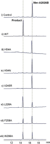
Activity measurements of PieE WT and selected mutants. Mer-A2026B is the substrate that is transformed into product after the reaction catalyzed by PieE. A standard PieE assay was performed in Tris-HCl buffer (50 mm, pH 8.0) containing 100 μm substrate, 5 μm enzyme (WT or mutant), 2 mm NADPH incubated at 25 °C for 2 h. The W296A mutant assay also included 50 μm FAD. For the control experiment, no enzyme was used. All assays were performed in single trials. The y axis (label omitted for clarity) shows the absorbance at 280 nm, and the x axis indicates the retention time in minutes.
Figure 6.
Figure 6.
Stereo view of the conformational changes induced by substrate binding. The path of signal transmission is indicated by the red arrows. Hydrogen bond interactions are shown in dashed lines. The substrate is represented in orange sticks. The binary complex structure is shown in gray with residues shown in pale green. The ternary complex structure is shown in yellow with residues colored in bright orange.
Figure 7.
Figure 7.
FAD adopting either IN or sliding conformation in the substrate-bound ternary complex. a, the FAD molecule is observed in the IN position in chain A and four other subunits. The chloride ion is shown as a magenta sphere. b, the FAD molecule adopts a unique sliding conformation in chain C of the ternary complex. In this binding mode, due to the change of orientation of Trp296, the interactions between Trp296 and FAD are significantly weaker as compared with the binary complex, where the Trp296 side chain and the isoalloxazine ring of FAD establish parallel π-π stacking interactions (Fig. 3). The N3 atom of the isoalloxazine ring establishes a hydrogen bond with the substrate molecule. Hydrogen bonds are shown in dashed lines. The Fourier difference maps (FoFc) with ligands omitted from refinement are shown in green mesh and contoured at 2.5σ.
Figure 8.
Figure 8.
Three distinct conformations of FAD in PieE. The color scheme is the same as in the previous figures. a, superposition of FAD OUT (carbon in gray) and FAD sliding conformation (carbon in yellow). In the sliding conformation, Trp296 (carbon in bright orange) undergoes a pronounced shift and establishes a hydrogen bond with Thr210, whereas the FAD molecule hydrogen-bonds to the substrate molecule. It should be noted that the shifted Trp296 (carbon in bright orange) and the sliding FAD are not parallel, thus preventing the formation of optimal π-π stacking interactions as observed for the OUT conformation of FAD in the binary complex (carbon of Trp296 in pale green). b, side view of superposition shown in a. c, superposition of OUT, sliding, and IN (carbon in cyan) conformations of FAD in PieE.
Scheme 1.
Scheme 1.
Schematic representation of the putative order of binding and chemical events in the catalytic cycle of PieE and other pertinent Group A flavin-dependent monooxygenases. The catalytic cycle is initiated by binding of the substrate, thus forming the enzyme-FAD-substrate complex. This event leads to the sliding conformation of FAD, followed by NADPH binding. Although it remains to be experimentally observed, upon NADPH binding, the oxidized FAD molecule most likely stays in the sliding conformation or in a very similar conformation (denoted as [SLIDING]). In this [sliding] conformation, oxidized FAD is reduced by NADPH, followed by the release of NADP+. These steps constitute the reductive half-reaction. In the subsequent oxidative half-reaction, the reduced FAD shifts to the IN conformation, followed by the reaction with molecular oxygen to form C4a-hydroperoxyflavin. The next step is the monooxygenation of substrate to yield the hydroxylated product along with the formation of C4a-hydroxyflavin. A dehydration reaction leads to the release of the final product and water. This last step terminates the cycle with regeneration of the oxidized FAD returning to the OUT conformation. Importantly, the mobile flavin alternates in at least three different conformations, namely OUT, sliding, and IN, to fulfill the catalytic cycle.
Figure 9.
Figure 9.
NADPH oxidation activity of WT PieE and the W296A mutant with the initial rates indicated. The activity of WT PieE was measured in 50 mm potassium phosphate buffer (pH 6.5) containing 100 μm FAD in the following conditions: substrate + NADPH (blue filled squares), substrate + 200 mm NaCl + NADPH (red filled circles), NADPH + 200 mm NaCl (green filled circles), and NADPH (black open squares). The autoxidation of NADPH (without PieE) is shown with gray open circles. The activity of the W296A mutant was measured in the same buffer containing FAD with substrate and NADPH (blue filled squares) and with NADPH but without substrate (black open squares). The enzyme (mutant and WT) concentration was set at 5 μm, whereas NADPH was set at 100 μm. The experiment was performed as a single trial.

Similar articles

Cited by

References

    1. Walsh C. T., and Wencewicz T. (2013) Flavoenzymes: versatile catalysts in biosynthetic pathways. Nat. Prod. Rep. 30, 175–200 10.1039/C2NP20069D - DOI - PMC - PubMed
    1. Ellis H. R. (2010) The FMN-dependent two-component monooxygenase systems. Arch. Biochem. Biophys. 497, 1–12 10.1016/j.abb.2010.02.007 - DOI - PubMed
    1. Chenprakhon P., Wongnate T., and Chaiyen P. (2019) Monooxygenation of aromatic compounds by flavin-dependent monooxygenases. Protein Sci. 28, 8–29 10.1002/pro.3525 - DOI - PMC - PubMed
    1. Teufel R., Agarwal V., and Moore B. S. (2016) Unusual flavoenzyme catalysis in marine bacteria. Curr. Opin. Chem. Biol. 31, 31–39 10.1016/j.cbpa.2016.01.001 - DOI - PMC - PubMed
    1. Huijbers M. M. E., Montersino S., Westphal A. H., Tischler D., and van Berkel W. J. H. (2014) Flavin dependent monooxygenases. Arch. Biochem. Biophys. 544, 2–17 10.1016/j.abb.2013.12.005 - DOI - PubMed

Publication types

MeSH terms

Associated data

Grants and funding

LinkOut - more resources