. 2020 Apr 3;295(14):4748-4759.
doi: 10.1074/jbc.RA119.010951. Epub 2020 Feb 28.

The microtubule-associated histone methyltransferase SET8, facilitated by transcription factor LSF, methylates α-tubulin

Affiliations

The microtubule-associated histone methyltransferase SET8, facilitated by transcription factor LSF, methylates α-tubulin

Hang Gyeong Chin et al. J Biol Chem. .

Abstract

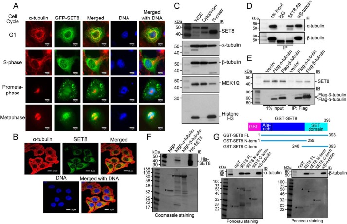
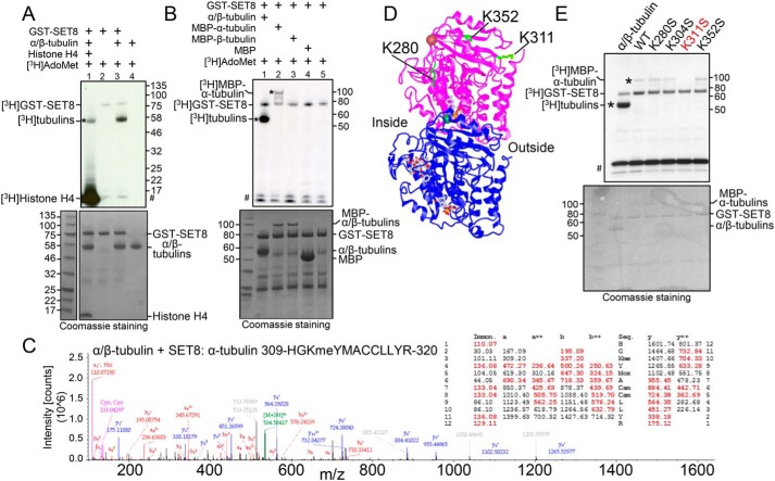
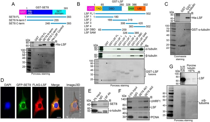
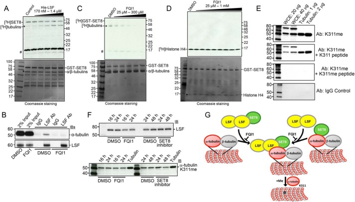
Microtubules are cytoskeletal structures critical for mitosis, cell motility, and protein and organelle transport and are a validated target for anticancer drugs. However, how tubulins are regulated and recruited to support these distinct cellular processes is incompletely understood. Posttranslational modifications of tubulins are proposed to regulate microtubule function and dynamics. Although many of these modifications have been investigated, only one prior study reports tubulin methylation and an enzyme responsible for this methylation. Here we used in vitro radiolabeling, MS, and immunoblotting approaches to monitor protein methylation and immunoprecipitation, immunofluorescence, and pulldown approaches to measure protein-protein interactions. We demonstrate that N-lysine methyltransferase 5A (KMT5A or SET8/PR-Set7), which methylates lysine 20 in histone H4, bound α-tubulin and methylated it at a specific lysine residue, Lys311 Furthermore, late SV40 factor (LSF)/CP2, a known transcription factor, bound both α-tubulin and SET8 and enhanced SET8-mediated α-tubulin methylation in vitro In addition, we found that the ability of LSF to facilitate this methylation is countered by factor quinolinone inhibitor 1 (FQI1), a specific small-molecule inhibitor of LSF. These findings suggest the general model that microtubule-associated proteins, including transcription factors, recruit or stimulate protein-modifying enzymes to target tubulins. Moreover, our results point to dual functions for SET8 and LSF not only in chromatin regulation but also in cytoskeletal modification.

Keywords: SET8; cancer; cytoskeleton; lysine methyltransferase 5A (KMT5A); mammalian cells; posttranslational modification (PTM); protein methylation; transcription factor CP2 (TFCP2); transcription factor LSF; tubulin.


Conflict of interest statement

The authors declare that they have no conflicts of interest with the contents of this article. The content is solely the responsibility of the authors and does not necessarily represent the official views of the National Institutes of Health

Figures

Figure 1.
Figure 1.
SET8 associates with tubulin in cells and directly interacts with α-tubulin in vitro. A, colocalization of SET8 and α-tubulin in COS7 cells. GFP-SET8 (green) was expressed in asynchronous cells, tubulin was detected with anti-α-tubulin antibody (red), and DNA with DAPI (blue). Yellow in the merged image indicates colocalization of SET8 and α-tubulin. Images are from cells identified as being in the indicated stages of cell cycle progression. Scale bars = 10 μm. B, colocalization of endogenous SET8 and α-tubulin in human HCT116 cells. C, endogenous SET8 is localized in the nucleus and cytoplasm in HEK293T cells. 10 μg each of whole-cell extract (WCE), cytoplasmic, and nuclear fractions were analyzed for the presence of SET8, α- and β-tubulins (cytoplasmic marker), MEK1/2 (predominantly cytoplasmic marker), and histone H3 (nuclear marker). D, coimmunoprecipitation from HEK293T cells of endogenous tubulins with endogenous SET8, using SET8 antibody (Ab). Right lane, more than 99% pure tubulin (MP Biomedicals, 08771121) as a positive control. Immunoprecipitates were analyzed using antibodies against the indicated proteins by immunoblot (IB). E, coimmunoprecipitation from HEK293T cells of endogenous SET8 with transiently expressed FLAG-tagged tubulins, as detected by immunoprecipitation (IP) with antibody against FLAG. F, MBP pulldown analysis of purified His-SET8 with MBP–α-tubulin but not MBP–β-tubulin. Top, IB in which biotinylated molecular mass markers are visualized. Bottom, Coomassie staining of the same gel (shown in grayscale) in which standard molecular mass markers are visualized. Asterisks, expected positions of migration of the MBPs. G, GST pulldown analysis of purified porcine brain tubulin to full length or the indicated overlapping segments of SET8 fused to GST. Top, IB in which biotinylated molecular mass markers are visualized. Bottom, Ponceau staining of the same gels (shown in grayscale) in which standard molecular mass markers are visualized. Asterisks, expected positions of migration of the GST proteins.
Figure 2.
Figure 2.
The histone methyltransferase SET8 methylates α-tubulin at Lys311. A, purified porcine tubulin (rPeptide, T-1201-1) is methylated by SET8. Lane 1, histone H4 (1 μg) was added in addition to tubulin as substrates. Top, autoradiogram of methyltransferase assays, showing methylation of tubulin (asterisk), histone H4, and automethylation of GST-SET8. #, migration of 3H-labeled impurities that migrated at a similar position as that of histone H4. Bottom, Coomassie staining of the same gel (shown in grayscale), indicating relative levels of the components in the reaction. B, recombinant human MBP–α-tubulin (asterisk), but not MBP–β-tubulin, is methylated by SET8. Autoradiogram (top) and Coomassie staining (bottom) are as described in A. Protein bands of less than 50 kDa are from the purified GST-SET8 preparation and are more evident in this experiment than in other reactions. C, mass spectrum and table of the expected m/z of the peptide fragments (with observed fragments in red), confirming methylation on Lys311 of α-tubulin after incubation of purified tubulin with SET8. D, the 3D structure of the α/β-tubulin heterodimer (PDB code 1JFF; purple, α-tubulin; blue, β-tubulin), indicating positions of lysines in α-tubulin targeted by SET8 in vitro (green). Inside and outside surfaces of the MT structure are indicated. E, mutation solely of Lys311 in recombinant MBP–α-tubulin (K311S) substantially reduced methylation by GST-SET8 in vitro. Autoradiogram (top) and Coomassie-staining (bottom) are as described in A.
Figure 3.
Figure 3.
LSF interacts directly with SET8 and tubulin. A, GST pulldown analysis of purified His-LSF to full length or the indicated overlapping segments of SET8 fused to GST. Top, IB in which biotinylated molecular mass markers are visualized. Bottom, Ponceau staining of the same gels (shown in grayscale) in which standard molecular mass markers are visualized. Asterisks, expected positions of migration of the GST proteins. B, GST pulldown analysis of purified porcine tubulin to purified full length or the indicated overlapping segments of LSF fused to GST. Gels are as described in A. Asterisks and bracket, expected positions of migration of the GST proteins. C, GST pulldown analysis of recombinant, purified His-tagged LSF to purified α-tubulin fused to GST. Gels are as described in A, except that the protein gel was stained with Coomassie. D, plasmids expressing 3×FLAG-LSF and GFP-SET8 were transfected into COS7 cells. Anti-FLAG antibody was visualized with a red fluorescent secondary antibody, and DNA was visualized with DAPI. The merged image indicates colocalization of GFP-SET8 with FLAG-LSF (yellow), concentrated largely near the nuclear membrane (Manders correlation coefficient of LSF and SET8 colocalization is 0.9, as determined via ImageJ 3D analysis). The majority of overexpressed 3×FLAG-LSF was cytoplasmic, with only a minority detected in the nucleus. E, specific coimmunoprecipitation of endogenous SET8 (top) and endogenous α-tubulin (bottom) from HEK293 cellular extracts, using antibodies to LSF compared with control IgG. F, specific coimmunoprecipitation of endogenous LSF from HEK293 cellular extracts using antibodies to SET8 (Ab1, Active Motif; Ab2, Millipore) compared with control IgG. Coimmunoprecipitation of PCNA and UHRF1 is also shown as a positive control. G, immunoblotting of purified porcine brain tubulin (rPeptide, >97%) shows the presence of LSF using an LSF mAb; representative also of results obtained using a separate source of purified tubulin: MP Biomedicals, more than 99%. Positive control for LSF migration, HEK293T whole-cell extract (293T WCE). Top, immunoblot. Bottom, Ponceau staining using standard molecular mass markers.
Figure 4.
Figure 4.
LSF and FQI1 oppositely affect methylation of tubulin by SET8. A, tubulin (>99%, MP Biomedicals) methylation reactions were performed with addition of the indicated increasing range of concentrations of LSF. Top, autoradiogram of methyltransferase assays, showing methylation of tubulin (asterisk) and automethylation of GST-SET8. The higher relative levels of GST-SET8 to α/β-tubulin in this experiment led to greater initial automethylation of SET8 relative to tubulin methylation. #, migration of 3H-labeled impurities. Bottom, Coomassie staining of the same gel (shown in grayscale), indicating relative levels of the components in the reaction. As in Fig. 2B, protein bands of less than 50 kDa are from the purified GST-SET8 preparation and are more evident in this experiment. B, coimmunoprecipitation of endogenous α-tubulin with endogenous LSF from HEK293T cell lysates was disrupted upon treatment of the cells with 2.5 μm FQI1 for 24 h. C, tubulin (>99%, MP Biomedicals) methylation reactions were performed with addition of the indicated increasing range of concentrations of FQI1. At 100 μm FQI1 (lane 4), methylation is decreased ∼3-fold. Gels are labeled as in A. D, histone H4 methylation reactions at limiting amounts of histone H4 (200 ng) were performed with addition of the indicated increasing range of concentrations of FQI1. Gels are labeled as in A. E, specific methylation of tubulin on Lys311. Immunoblots of HEK293T cell lysates and purified tubulin, at the indicated concentrations, with α-tubulin K311me or nonspecific IgG antibodies. Specificity to methylated Lys311 was demonstrated by preincubation of the antibody with methylated versus nonmethylated α-tubulin Lys311 peptides. F, treatment of HEK293T cells with either LSF or SET8 inhibitors somewhat reduces the level of methylated Lys311 on α-tubulin. G, model of recruitment and/or activation of SET8 at microtubules by LSF and subsequent methylation of α-tubulin by SET8.

References

    1. Verhey K. J., and Gaertig J. (2007) The tubulin code. Cell Cycle 6, 2152–2160 10.4161/cc.6.17.4633 - DOI - PubMed
    1. Janke C. (2014) The tubulin code: molecular components, readout mechanisms, and functions. J. Cell Biol. 206, 461–472 10.1083/jcb.201406055 - DOI - PMC - PubMed
    1. Song Y., and Brady S. T. (2015) Post-translational modifications of tubulin: pathways to functional diversity of microtubules. Trends Cell Biol. 25, 125–136 10.1016/j.tcb.2014.10.004 - DOI - PMC - PubMed
    1. Wloga D., and Gaertig J. (2010) Post-translational modifications of microtubules. J. Cell Sci. 123, 3447–3455 10.1242/jcs.063727 - DOI - PMC - PubMed
    1. Janke C., and Montagnac G. (2017) Causes and consequences of microtubule acetylation. Curr. Biol. 27, R1287–R1292 10.1016/j.cub.2017.10.044 - DOI - PubMed

Publication types

Associated data

LinkOut - more resources