. 2020 Feb 28;11(1):1064.
doi: 10.1038/s41467-020-14344-7.

Inhalation of lung spheroid cell secretome and exosomes promotes lung repair in pulmonary fibrosis

Affiliations

Inhalation of lung spheroid cell secretome and exosomes promotes lung repair in pulmonary fibrosis

Phuong-Uyen C Dinh et al. Nat Commun. .

Abstract

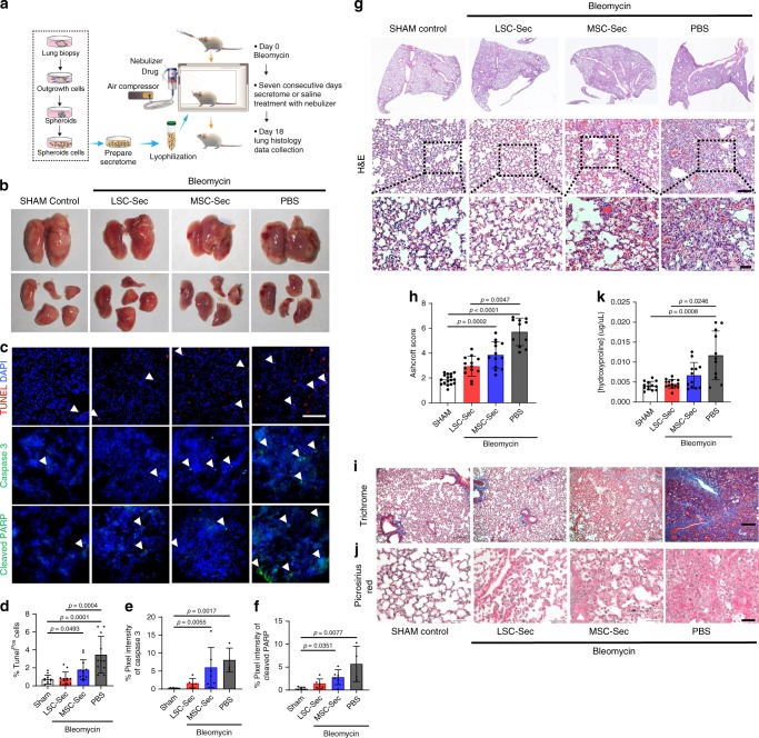
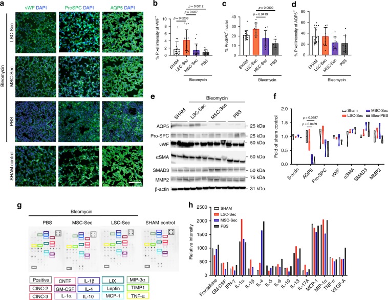
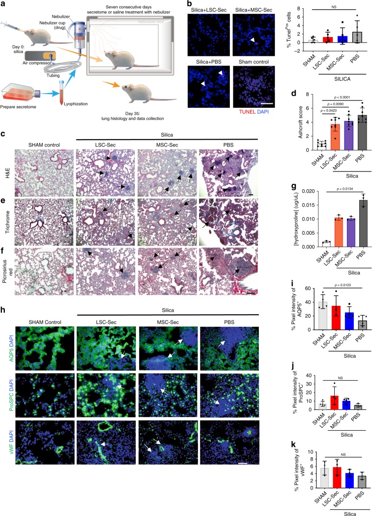
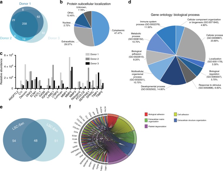
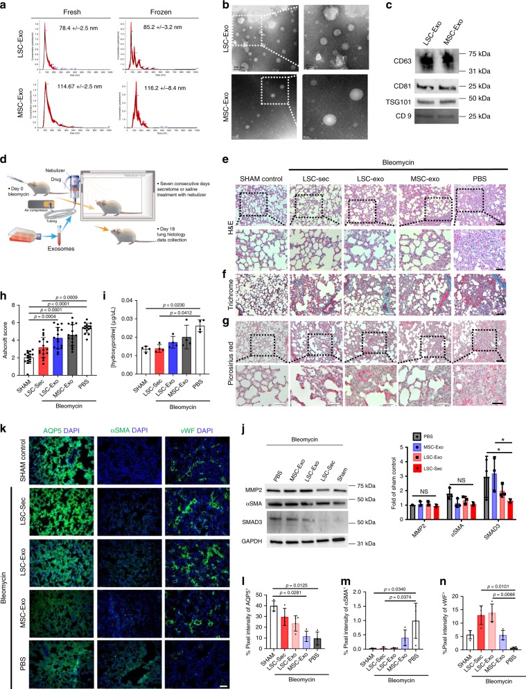
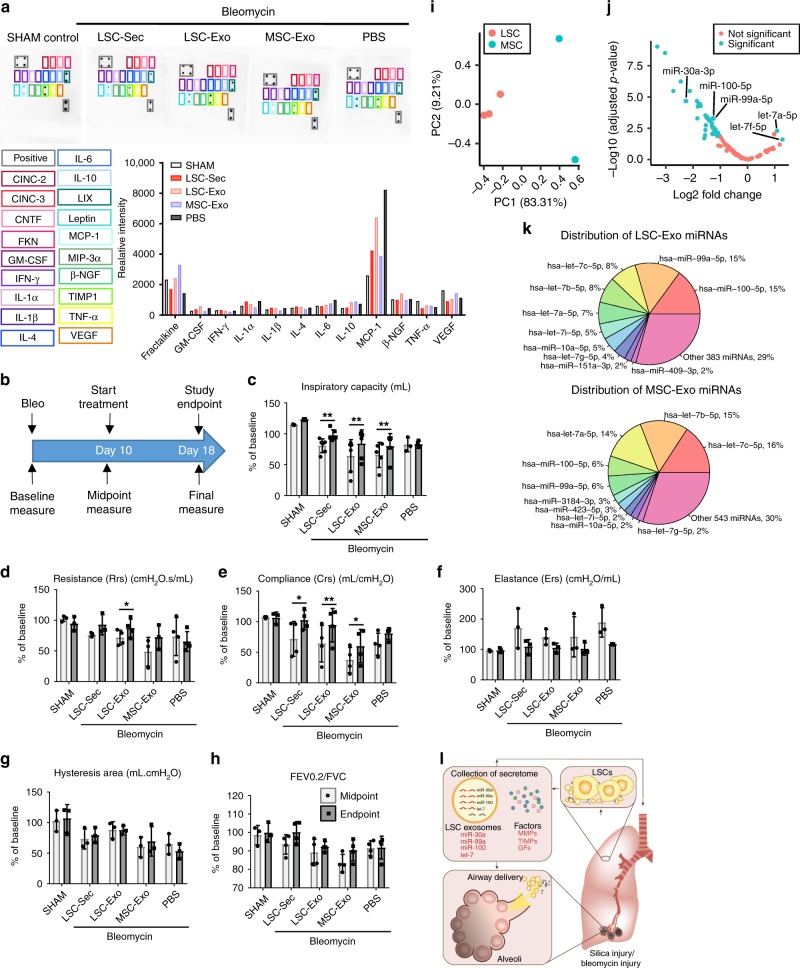
Idiopathic pulmonary fibrosis (IPF) is a fatal and incurable form of interstitial lung disease in which persistent injury results in scar tissue formation. As fibrosis thickens, the lung tissue loses the ability to facilitate gas exchange and provide cells with needed oxygen. Currently, IPF has few treatment options and no effective therapies, aside from lung transplant. Here we present a series of studies utilizing lung spheroid cell-secretome (LSC-Sec) and exosomes (LSC-Exo) by inhalation to treat different models of lung injury and fibrosis. Analysis reveals that LSC-Sec and LSC-Exo treatments could attenuate and resolve bleomycin- and silica-induced fibrosis by reestablishing normal alveolar structure and decreasing both collagen accumulation and myofibroblast proliferation. Additionally, LSC-Sec and LSC-Exo exhibit superior therapeutic benefits than their counterparts derived from mesenchymal stem cells in some measures. We showed that an inhalation treatment of secretome and exosome exhibited therapeutic potential for lung regeneration in two experimental models of pulmonary fibrosis.


Conflict of interest statement

K.C. is a co-founder and equity holder of BreStem Therapeutics. BreStem provided no funding to this study. The remaining authors declare no competing interests.

Figures

Fig. 1
Fig. 1. LSC-Sec inhalation reverses alveolar epithelial cell damage caused by Bleomycin injury.
a The schematic of LSC-Sec procedure and study design; n = 12 biological independent animals per group. b Macroscopic view of the explanted lungs at study endpoint. c Representative TUNEL (top), Caspase 3 (middle), and cleaved PARP (bottom) immunostaining of apoptotic cells for each treatment group. df Quantification of percent of Tunel (d) n = 12 biological independent animals, Caspase 3 (e) n = 6 biological independent animals, and cleaved PARP (f) positive cells, n = 6 biological independent animals; Scale bar = 100 μm; each dot represents data from one animal; n = 6–12. g Representative H&E staining. Top: Scale bar = 500 μm Bottom: Scale bar = 200 μm. h Quantification of fibrosis by Ashcroft score; each dot represents data from one animal; n = 12 biological independent animals. Ashcroft score was performed by averaging the score from two blinded and one non-blinded scorer. i Representative Gomori’s trichrome staining; muscle fibers (red), collagen (blue), nuclei (black-purple), and erythrocytes (red). Scale bar = 1000 μm. j Representative picrosirius red staining; Collagen types I and III (red). Scale bar = 50 μm. k Quantification of pulmonary hydroxyproline levels; each dot represents data from one animal; n = 12 biological independent animals. Throughout, data are mean ± s.d. P-value as indicated by non-parametric one-way ANOVA.
Fig. 2
Fig. 2. LSC-Sec inhalation treatment promotes alveolar repair.
a Representative immunostaining of lung sections for von Willebrand Factor (vWF), pro-surfactant protein C (Pro-SPC) and aquaporin 5 (AQP5). Scale bar = 100 μm. bd Quantification of percent pixel intensity of vWF+ (b), percent ProSPC+ nuclei (c), and percent pixel intensity of AQP5+ (d); each dot represents data from one animal; n = 12 biological independent animals. ef Immunoblot analysis of aquaporin 5 (AQP5), pro-surfactant protein C (Pro-SPC), von Willebrand Factor (vWF), alpha smooth muscle actin (αSMA), SMAD3, matrix metalloproteinase 2 (MMP-2), and beta-actin loading control (B-actin) from lung protein lysate (e) with corresponding quantification of protein levels as fold of sham control (f); each dot represents data from one animal; n = 3 biological independent animals. gh Representative cytokine array with quantification of relative intensity (g) and corresponding quantification of relative intensity (h). Throughout, data are mean ± s.d. P-value as indicated by non-parametric one-way ANOVA.
Fig. 3
Fig. 3. Lung repair and fibrosis in mice after silica-injury.
a The experimental study schematic of the silica-induced fibrosis study in A/J mice; n = 10 biological independent animals per group. b Representative TUNEL staining of apoptotic cells for each treatment group and quantification of percent of Tunel positive cells; Scale bar = 50 μm; each dot represents data from one animal; n = 5 biological independent animals. c Representative H&E staining. Scale bar = 100 μm. d Quantification of fibrosis by Ashcroft score; each dot represents data from one animal; n = 8 biological independent animals. Ashcroft score was performed by averaging the score from two blinded and one non-blinded scorer. e Gomori’s trichrome staining: muscle fibers (red), collagen (blue), nuclei (black-purple) and erythrocytes (red). Scale bar = 100 μm. f Representative picrosirius red staining; Collagen types I and III (red). Scale bar = 100 μm. g Quantification of pulmonary hydroxyproline levels; each dot represents data from one animal; n = 4 biological independent animals. h Representative immunostaining of lung sections for aquaporin 5 (AQP5), pro-surfactant protein C (Pro-SPC) and von Willebrand Factor (vWF) Scale bar = 50 μm. ik Quantification of percent pixel intensity of AQP5+ (i), ProSPC+ (j), and vWF+ (k). Each dot represents data from one animal; n = 4 biological independent animals. Throughout, data are mean ± s.d. P-value as indicated by non-parametric one-way ANOVA; NS, not significant.
Fig. 4
Fig. 4. Proteomic analysis of LSC-Sec and LSC-Exo.
a Venn diagram of proteins identified in LSC-Sec derived from three donors. b Pie chart of protein subcellular location of all common proteins in all three LSC-Sec. c Relative abundance of 20 of the 103 extracellular proteins identified in all three LSC-Sec. d Gene ontology pie chart of biological process associated with the shared extracellular proteins. e Venn diagram of extracellular proteins identified in LSC-Sec versus LSC-Exo. f Gene ontology chord chart of biology process associated with the shared LSC-Exo proteins.
Fig. 5
Fig. 5. Therapeutic potential of exosome inhalation treatment in rats with pulmonary fibrosis.
a Size analysis of fresh, frozen and lyophilized exosome particles by NanoSight. b Representative transmission electron micrograph (TEM) of LSC-Exo. Left scale bar = 0.2 μm. Right scale bar = 50 nm. c Immunoblot analysis of CD63, CD81, TSG101, and CD9 protein in LSC-Exo and MSC-Exo. d The experimental study schematic of the exosome study in SD rats; n = 12 biological independent animals per group. e Representative H&E staining Top: Scale bar = 100 μm Bottom: Scale bar = 50 μm. f Representative Gomori’s trichrome staining Scale bar = 100 μm (bottom); muscle fibers (red), collagen (blue), nuclei (black-purple) and erythrocytes (red). g Representative picrosirius red staining; Collagen types I and III (red); Scale bar = 50 μm. h Quantification of fibrosis by Ashcroft score; Each dot represents data from one animal; n = 12 biological independent animals. Ashcroft score was performed by averaging the score from one blinded and one non-blinded scorer. i Quantification of pulmonary hydroxyproline levels; *P ≤ 0.05; each dot represents data from one animal; n = 4 biological independent animals. j Immunoblot analysis of matrix metalloproteinase 2 (MMP-2), alpha smooth muscle actin (αSMA), SMAD3 and GAPDH loading control. k Representative immunostaining of lung sections for aquaporin 5 (AQP5), alpha-smooth muscle actin (αSMA), and von Willebrand Factor (vWF). Scale bar = 75 μm. ln Quantification of pixel intensity of AQP5 (l), α-SMA (m), and vWF (n). Each dot represents data from one animal; n = 4 biological independent animals. Throughout, data are mean ± s.d. P-value as indicated by non-parametric one-way ANOVA.
Fig. 6
Fig. 6. Exosome treatment improves pulmonary function post-Bleo and exosome miRNA profiling.
a Representative rat cytokine array detecting 19 rat proteins from blood serum. b The schematic of pulmonary function measurements. cg Quantification of lung function measurements (c) pulmonary inspiratory capacity; (d) pulmonary elastance (e) pulmonary resistance; (f) pulmonary compliance; (g) hysteresis area; (h) forced expiratory volume (FEV) to forced vital capacity (FVC) ratio. i Principal component analysis chart of LSC-exosome and MSC-exosome microRNA content. j Volcano plot of differentially expressed miRNA of LSC-exosomes and MSC-exosomes miRNA content. k Distribution of the top 10 miRNAs in LSC-exosomes (top) and MSC-exosomes (bottom). l LSC secreted exosomes and soluble factors modulate alveolar repair and fibrosis. Throughout, data are mean ± s.d. P-value as indicated by non-parametric one-way ANOVA (a) and Student’s paired t-test (ch).

References

    1. Henry Eric, Cores Jhon, Hensley M. Taylor, Anthony Shirena, Vandergriff Adam, de Andrade James B.M., Allen Tyler, Caranasos Thomas G., Lobo Leonard J., Cheng Ke. Adult Lung Spheroid Cells Contain Progenitor Cells and Mediate Regeneration in Rodents With Bleomycin-Induced Pulmonary Fibrosis. STEM CELLS Translational Medicine. 2015;4(11):1265–1274. doi: 10.5966/sctm.2015-0062. - DOI - PMC - PubMed
    1. Dinh, P. C. et al. Derivation of therapeutic lung spheroid cells from minimally invasive transbronchial pulmonary biopsies. Respir. Res.18, 132 (2017). - PMC - PubMed
    1. Cores J, et al. Safety and efficacy of allogeneic lung spheroid cells in a mismatched rat model of pulmonary fibrosis. Stem Cells Transl. Med. 2017;6:1905–1916. doi: 10.1002/sctm.16-0374. - DOI - PMC - PubMed
    1. Yang, H. et al. Neural stem cell-conditioned medium ameliorated cerebral ischemia-reperfusion injury in rats. Stem Cells Int. 2018, 4659159 (2018). - PMC - PubMed
    1. Bai L, et al. Hepatocyte growth factor mediates mesenchymal stem cell-induced recovery in multiple sclerosis models. Nat. Neurosci. 2012;15:862–870. doi: 10.1038/nn.3109. - DOI - PMC - PubMed

Publication types

MeSH terms