. 2020 Feb 28;10(1):3699.
doi: 10.1038/s41598-020-60631-0.

Insight into DNA substrate specificity of PARP1-catalysed DNA poly(ADP-ribosyl)ation

Affiliations

Insight into DNA substrate specificity of PARP1-catalysed DNA poly(ADP-ribosyl)ation

Elie Matta et al. Sci Rep. .

Abstract

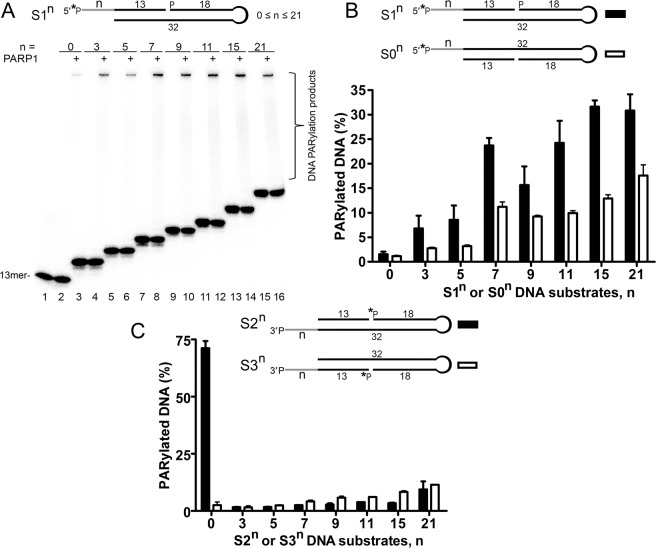
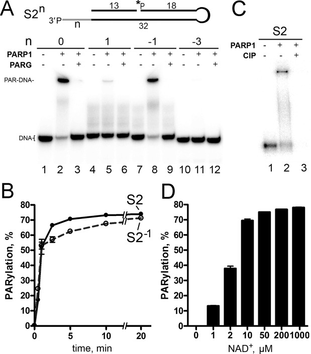
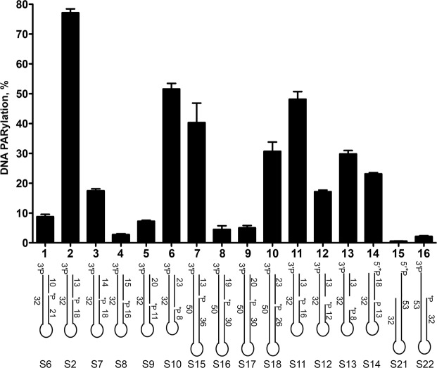
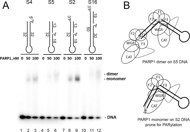
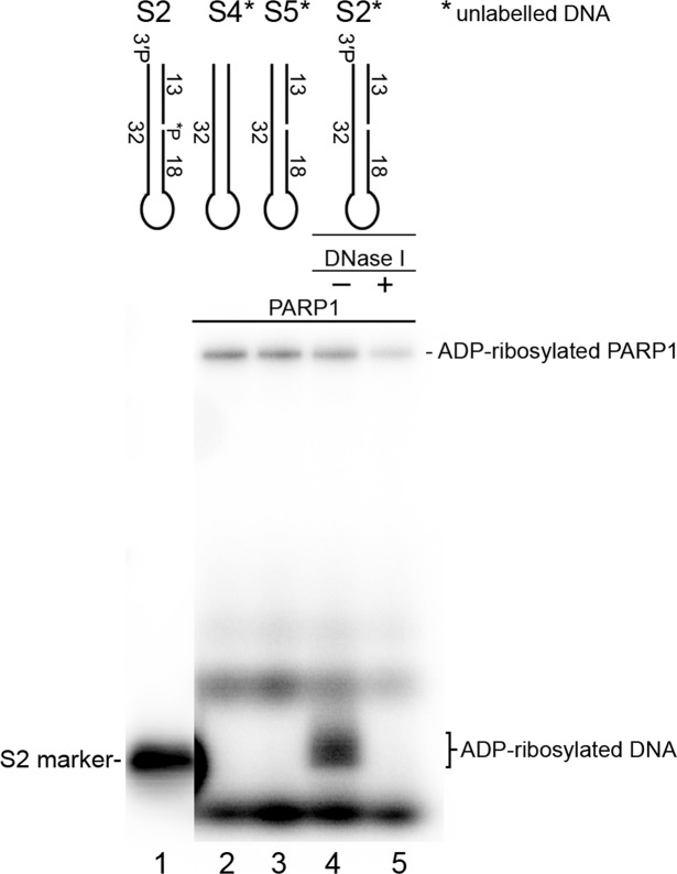
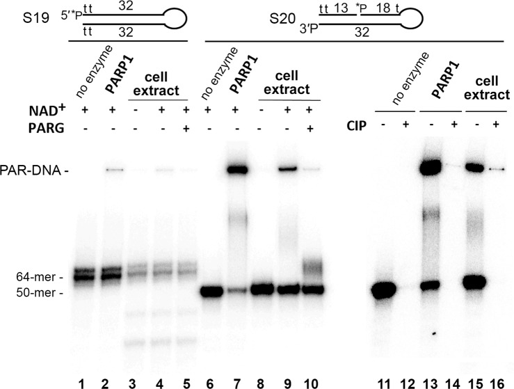
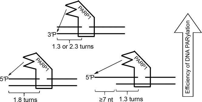
DNA-dependent poly(ADP-ribose) polymerases (PARPs) PARP1, PARP2 and PARP3 act as DNA break sensors signalling DNA damage. Upon detecting DNA damage, these PARPs use nicotine adenine dinucleotide as a substrate to synthesise a monomer or polymer of ADP-ribose (MAR or PAR, respectively) covalently attached to the acceptor residue of target proteins. Recently, it was demonstrated that PARP1-3 proteins can directly ADP-ribosylate DNA breaks by attaching MAR and PAR moieties to terminal phosphates. Nevertheless, little is still known about the mechanisms governing substrate recognition and specificity of PARP1, which accounts for most of cellular PARylation activity. Here, we characterised PARP1-mediated DNA PARylation of DNA duplexes containing various types of breaks at different positions. The 3'-terminal phosphate residue at double-strand DNA break ends served as a major acceptor site for PARP1-catalysed PARylation depending on the orientation and distance between DNA strand breaks in a single DNA molecule. A preference for ADP-ribosylation of DNA molecules containing 3'-terminal phosphate over PARP1 auto-ADP-ribosylation was observed, and a model of DNA modification by PARP1 was proposed. Similar results were obtained with purified recombinant PARP1 and HeLa cell-free extracts. Thus, the biological effects of PARP-mediated ADP-ribosylation may strongly depend on the configuration of complex DNA strand breaks.


Conflict of interest statement

The authors declare no competing interests.

Figures

Figure 1
Figure 1
Effects of the type and size of protruding ends in Dbait-based DNA structures containing a 1-nt gap on the PARP1-catalysed formation of PAR–DNA adducts. Twenty-nanomolar PARP1 was incubated with 20 nM 5′-[32P]labelled oligonucleotide and 1 mM NAD+ for 15 min at 37 °C under standard reaction conditions. (A) Denaturing PAGE analysis of PARP1-generated products of PARylation of [32P]labelled DNA substrates S1n. (B) and (C) Comparison of DNA PARylation activities of PARP1 towards DNA substrates containing 5′-overhangs (S0n and S1n) or 3′-overhangs (S2n and S3n), respectively. The data on PARP-catalysed formation of PAR-DNA products are presented as mean ± SD from three independent experiments.
Figure 2
Figure 2
PARP1-catalysed PARylation of S2n DNA structures with a 3′-phosphate terminus at the DBS end. (A) [32P]labelled DNA substrates S2n (50 nM) were incubated with 40 nM PARP1 for 10 min 37 °C. (B) Time dependence of PARP1-driven PARylation of substrates S2 and S2-1. DNA substrates (50 nM) were incubated with 20 nM PARP1 for the indicated period under standard reaction conditions. (C) CIP-induced dephosphorylation of 5′-[32P]labelled PAR-S2 products. After incubation with PARP1, the S2 samples were heated for 10 min at 85 °C, and the resulting [32P]labelled DNA PARylation products were further incubated with 10 U of CIP for 30 min at 37 °C. (D) The dependence of S2 DNA (40 nM) PARylation by PARP1 (20 nM) on NAD+ concentration. The data in panels C and D are presented as mean ± SD from three independent experiments.
Figure 3
Figure 3
Gap–DSB distance dependence of PARP1-catalysed PARylation of the 3′ phosphate at a DSB end of DNA duplexes. The data on PARP-catalysed formation of PAR-DNA products are presented as mean ± SD from three independent experiments performed under standard reaction conditions.
Figure 4
Figure 4
The monomeric mode of PARP1 binding to DNA molecules prone to PARylation. (A) The EMSA. Each of 20 nM DNA duplexes was incubated with 0, 50 or 100 nM PARP1 in a buffer consisting of 20 mM Tris-HCl pH 7.6, 50 mM KCl and 1 mM DTT for 10 min at room temperature. The DNA–protein complexes were analysed by electrophoresis in a 4–12% Tris-Glycine polyacrylamide gel (Novex) under non-denaturing conditions at 4 °C after addition of 10% glycerol. (B) The putative model of PARP1 complexes with DNA substrates prone (S2) or not prone (S5) to DNA break PARylation.
Figure 5
Figure 5
Comparison of the efficiency of PARP1-catalysed auto- and DNA ADP-ribosylation. The denaturing SDS-PAGE analysis of the products of PARP1 incubation with cold oligonucleotide duplexes in the presence of [adenylate-32P]NAD+. For details see Materials and Methods.
Figure 6
Figure 6
Formation of PAR–3′ phosphate–DNA adducts in nuclear extracts from HeLa PARGKD cells. Fifty-nanomolar [32P]labelled S19 or S20 Dbait-based molecules were incubated with 2.5 µg/µl HeLa extracts or 40 nM PARP1 in the presence of 67 mM KCl, 10 mM HEPES-KOH pH 8.0 and 500 µM NAD+ for 20 min at 37 °C. The reactions were stopped by heating the samples for 10 min at 80 °C, and the resulting DNA PARylation products were next incubated with 20 pg/µl PARG (lanes 5 and 10) or after phenol-chloroform extraction with 10 U of CIP for 30 min at 37 °C (lanes 12, 14 and 16). The reaction products were analysed by denaturing PAGE.
Figure 7
Figure 7
Schematic representation of the putative model of DNA modification by PARP1 activated on a 1-nt gap.

References

    1. Mortusewicz O, Ame JC, Schreiber V, Leonhardt H. Feedback-regulated poly(ADP-ribosyl)ation by PARP-1 is required for rapid response to DNA damage in living cells. Nucleic acids research. 2007;35:7665–7675. doi: 10.1093/nar/gkm933. - DOI - PMC - PubMed
    1. Boehler C, et al. Poly(ADP-ribose) polymerase 3 (PARP3), a newcomer in cellular response to DNA damage and mitotic progression. Proceedings of the National Academy of Sciences of the United States of America. 2011;108:2783–2788. doi: 10.1073/pnas.1016574108. - DOI - PMC - PubMed
    1. Haince JF, et al. PARP1-dependent kinetics of recruitment of MRE11 and NBS1 proteins to multiple DNA damage sites. The Journal of biological chemistry. 2008;283:1197–1208. doi: 10.1074/jbc.M706734200. - DOI - PubMed
    1. Martin-Hernandez K, Rodriguez-Vargas JM, Schreiber V, Dantzer F. Expanding functions of ADP-ribosylation in the maintenance of genome integrity. Seminars in cell & developmental biology. 2017;63:92–101. doi: 10.1016/j.semcdb.2016.09.009. - DOI - PubMed
    1. Wei H, Yu X. Functions of PARylation in DNA Damage Repair Pathways. Genomics, proteomics & bioinformatics. 2016;14:131–139. doi: 10.1016/j.gpb.2016.05.001. - DOI - PMC - PubMed

Publication types