

Reconstitution of Membrane Proteins into Platforms Suitable for Biophysical and Structural Analyses
- PMID: 32112324
- PMCID: PMC9288841
- DOI: 10.1007/978-1-0716-0373-4_14
Reconstitution of Membrane Proteins into Platforms Suitable for Biophysical and Structural Analyses
Abstract
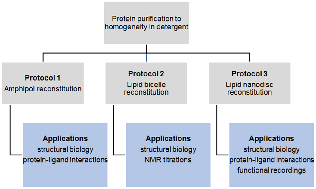
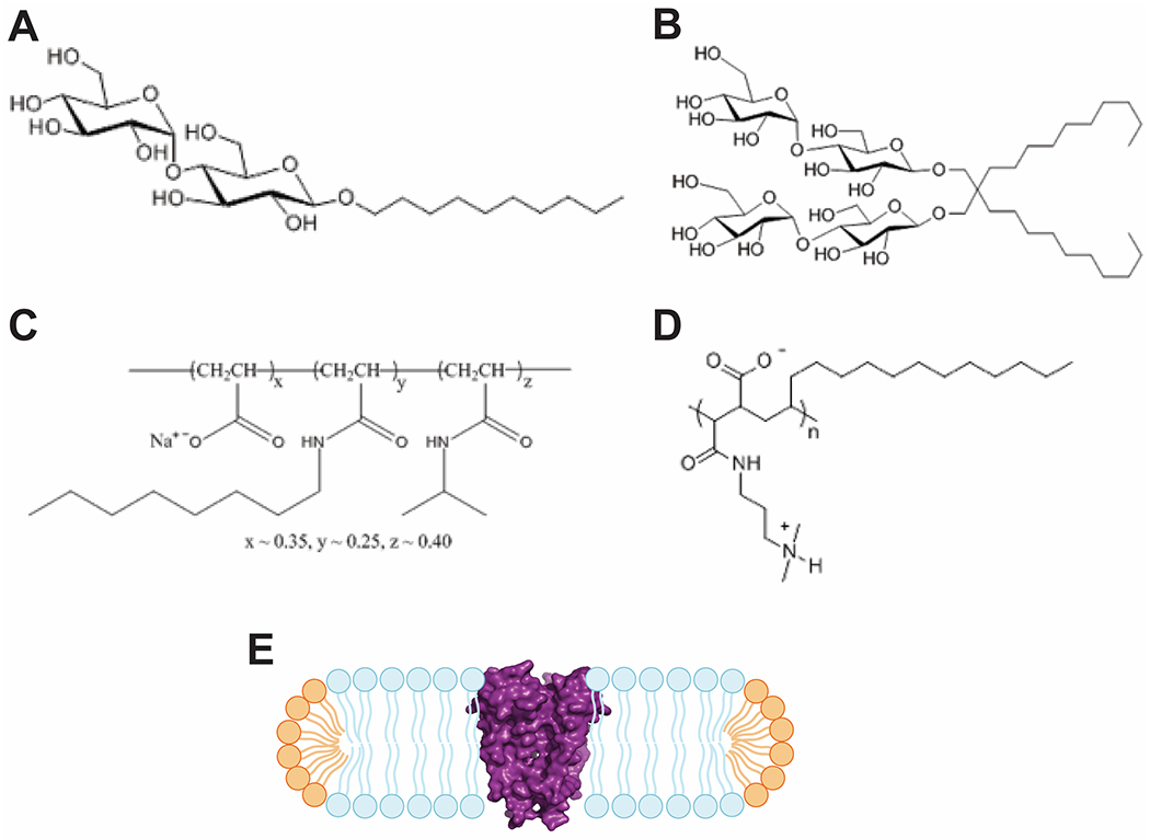
Integral membrane proteins have historically been challenging targets for biophysical research due to their low solubility in aqueous solution. Their importance for chemical and electrical signaling between cells, however, makes them fascinating targets for investigators interested in the regulation of cellular and physiological processes. Since membrane proteins shunt the barrier imposed by the cell membrane, they also serve as entry points for drugs, adding pharmaceutical research and development to the interests. In recent years, detailed understanding of membrane protein function has significantly increased due to high-resolution structural information obtained from single-particle cryo-EM, X-ray crystallography, and NMR. In order to further advance our mechanistic understanding on membrane proteins as well as foster drug development, it is crucial to generate more biophysical and functional data on these proteins under defined conditions. To that end, different techniques have been developed to stabilize integral membrane proteins in native-like environments that allow both structural and biophysical investigations-amphipols, lipid bicelles, and lipid nanodiscs. In this chapter, we provide detailed protocols for the reconstitution of membrane proteins according to these three techniques. We also outline some of the possible applications of each technique and discuss their advantages and possible caveats.
Keywords: Amphipol; Bicelles; Lipids; Membrane protein biophysics; Membrane proteins; Membrane scaffold; Nanodisc; Reconstitution.
Figures
Similar articles
-
Interfacing Membrane Mimetics with Mass Spectrometry.Acc Chem Res. 2016 Nov 15;49(11):2459-2467. doi: 10.1021/acs.accounts.6b00379. Epub 2016 Oct 13. Acc Chem Res. 2016. PMID: 27736086 Free PMC article.
-
Fake It 'Till You Make It-The Pursuit of Suitable Membrane Mimetics for Membrane Protein Biophysics.Int J Mol Sci. 2020 Dec 23;22(1):50. doi: 10.3390/ijms22010050. Int J Mol Sci. 2020. PMID: 33374526 Free PMC article. Review.
-
Membrane Protein Production in E. coli Lysates in Presence of Preassembled Nanodiscs.Methods Mol Biol. 2017;1586:291-312. doi: 10.1007/978-1-4939-6887-9_19. Methods Mol Biol. 2017. PMID: 28470613
-
Solubilization of Membrane Proteins into Functional Lipid-Bilayer Nanodiscs Using a Diisobutylene/Maleic Acid Copolymer.Angew Chem Int Ed Engl. 2017 Feb 6;56(7):1919-1924. doi: 10.1002/anie.201610778. Epub 2017 Jan 12. Angew Chem Int Ed Engl. 2017. PMID: 28079955 Free PMC article.
-
Nanodiscs for Structural Biology in a Membranous Environment.Chem Pharm Bull (Tokyo). 2019;67(4):321-326. doi: 10.1248/cpb.c18-00941. Chem Pharm Bull (Tokyo). 2019. PMID: 30930435 Review.
Cited by
-
Lipid Membrane Mimetics in Functional and Structural Studies of Integral Membrane Proteins.Membranes (Basel). 2021 Sep 3;11(9):685. doi: 10.3390/membranes11090685. Membranes (Basel). 2021. PMID: 34564502 Free PMC article. Review.
-
Correlating ion channel structure and function.Methods Enzymol. 2021;652:3-30. doi: 10.1016/bs.mie.2021.02.016. Epub 2021 Mar 25. Methods Enzymol. 2021. PMID: 34059287 Free PMC article.
References
-
- Fagerberg L, Jonasson K, von Heijne G, Uhlén M, Berglund L, Prediction of the human membrane proteome, Proteomics 10(6) (2010) 1141–9. - PubMed
Publication types
MeSH terms
Substances
Grants and funding
LinkOut - more resources
Full Text Sources
