The ratio of superoxide dismutase to standard deviation of erythrocyte distribution width as a predictor of systemic lupus erythematosus
- PMID: 32112599
- PMCID: PMC7307334
- DOI: 10.1002/jcla.23230
The ratio of superoxide dismutase to standard deviation of erythrocyte distribution width as a predictor of systemic lupus erythematosus
Abstract
Background: To explore the clinical value of the serum superoxide dismutase-to-standard deviation of erythrocyte distribution width ratio (SRSR) in systemic lupus erythematosus (SLE).
Methods: A total of 222 SLE patients from the Rheumatology and Immunology Department in the Second Affiliated Hospital of Chongqing Medical University from January 2017 to April 2019 were collected as the experimental group, and a total of 202 healthy physical examiners were extracted as the control group. Neutrophil-to-lymphocyte ratio (NLR), superoxide dismutase-to-standard deviation of erythrocyte distribution width ratio (SRSR), and platelet-to-lymphocyte ratio (PLR) were calculated from the collected data and then compared the level of the above three indexes between the two groups. In addition, we analyzed the association between SRSR and clinically relevant indicators.
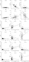
Results: We found that the SRSR of SLE patients was significantly lower than healthy control group, by analyzing the receiver operating characteristic (ROC) curve; it revealed that the SRSR had higher specificity and sensitivity than either superoxide dismutase (SOD) or standard deviation of erythrocyte distribution width (RDW-SD) alone. The area under the curve (AUC) for SRSR was significantly larger than either SOD or RDW-SD alone, and the AUC for SRSR was also larger than NLR and PLR. And it was found that SRSR was independently correlated with SLE disease activity through multiple linear regression analysis.
Conclusion: SRSR is a useful biomarker for the diagnosis of SLE, and it is of great significance in the clinical application.
Keywords: predictor; ratio; standard deviation of erythrocyte distribution width; superoxide dismutase; systemic lupus erythematosus.
© 2020 The Authors. Journal of Clinical Laboratory Analysis Published by Wiley Periodicals, Inc.
Figures


Similar articles
-
Neutrophil to lymphocyte ratio (NLR) and platelet to lymphocyte ratio (PLR) were useful markers in assessment of inflammatory response and disease activity in SLE patients.Mod Rheumatol. 2016;26(3):372-6. doi: 10.3109/14397595.2015.1091136. Epub 2016 Mar 4. Mod Rheumatol. 2016. PMID: 26403379
-
Hematological factors associated with immunity, inflammation, and metabolism in patients with systemic lupus erythematosus: Data from a Zhuang cohort in Southwest China.J Clin Lab Anal. 2020 Jun;34(6):e23211. doi: 10.1002/jcla.23211. Epub 2020 Jan 24. J Clin Lab Anal. 2020. PMID: 31978275 Free PMC article.
-
Clinical Usefulness of Hematologic Indices as Predictive Parameters for Systemic Lupus Erythematosus.Lab Med. 2020 Sep 1;51(5):519-528. doi: 10.1093/labmed/lmaa002. Lab Med. 2020. PMID: 32073127
-
Neutrophil to lymphocyte ratio and platelet to lymphocyte ratio in patients with systemic lupus erythematosus and their correlation with activity: A meta-analysis.Int Immunopharmacol. 2019 Nov;76:105949. doi: 10.1016/j.intimp.2019.105949. Epub 2019 Oct 18. Int Immunopharmacol. 2019. PMID: 31634817
-
Circulating antioxidant levels in systemic lupus erythematosus patients: a systematic review and meta-analysis.Biomark Med. 2019 Sep;13(13):1137-1152. doi: 10.2217/bmm-2019-0034. Epub 2019 Sep 2. Biomark Med. 2019. PMID: 31475863
Cited by
-
A systematic review and meta-analysis of the diagnostic accuracy of the neutrophil-to-lymphocyte ratio and the platelet-to-lymphocyte ratio in systemic lupus erythematosus.Clin Exp Med. 2024 Jul 25;24(1):170. doi: 10.1007/s10238-024-01438-5. Clin Exp Med. 2024. PMID: 39052098 Free PMC article.
-
Redox Homeostasis Involvement in the Pharmacological Effects of Metformin in Systemic Lupus Erythematosus.Antioxid Redox Signal. 2022 Mar;36(7-9):462-479. doi: 10.1089/ars.2021.0070. Epub 2022 Jan 4. Antioxid Redox Signal. 2022. PMID: 34619975 Free PMC article. Review.
-
A predictive model using risk factor categories for hospital-acquired pneumonia in patients with aneurysmal subarachnoid hemorrhage.Front Neurol. 2022 Dec 6;13:1034313. doi: 10.3389/fneur.2022.1034313. eCollection 2022. Front Neurol. 2022. PMID: 36561302 Free PMC article.
-
Measurement of superoxide dismutase: clinical usefulness for patients with anti-neutrophil cytoplasmic antibody-associated vasculitis.Adv Rheumatol. 2023 Jun 28;63(1):28. doi: 10.1186/s42358-023-00312-4. Adv Rheumatol. 2023. PMID: 37381048
-
The Role of the Oxidative State and Innate Immunity Mediated by TLR7 and TLR9 in Lupus Nephritis.Int J Mol Sci. 2023 Oct 16;24(20):15234. doi: 10.3390/ijms242015234. Int J Mol Sci. 2023. PMID: 37894915 Free PMC article. Review.
References
-
- Liao YQ, Xia HJ, Peng KJ, Liu JR. Correlation between oxidative reduction state changes and lymphocyte apoptosis and disease activity in systemic lupus erythematosus. Chin J Immunol. 2013;29:1288‐1292.
-
- Qun Z, Qing YD. Study on the correlation between lipid and protein peroxidation and clinical indexes of systemic lupus erythematosus. Chin J Rheumatol. 2008;12:95‐97.
-
- Guidelines for referral and management of systemic lupus erythematosus in adults. American College of Rheumatology Ad Hoc Committee on Systemic Lupus Erythematosus Guidelines. Arthritis Rheum. 1999;42:1785‐1796. - PubMed
-
- Gladman DD, Dominique L, Urowitz MB. Systemic lupus erythematosus disease activity index 2000. J Rheumatol. 2002;29(2):288‐291. - PubMed
MeSH terms
Substances
Grants and funding
LinkOut - more resources
Full Text Sources
Medical
Miscellaneous

