. 2020 Feb 29;17(1):75.
doi: 10.1186/s12974-019-1616-z.

The spinal microglial IL-10/β-endorphin pathway accounts for cinobufagin-induced mechanical antiallodynia in bone cancer pain following activation of α7-nicotinic acetylcholine receptors

Affiliations

The spinal microglial IL-10/β-endorphin pathway accounts for cinobufagin-induced mechanical antiallodynia in bone cancer pain following activation of α7-nicotinic acetylcholine receptors

Evhy Apryani et al. J Neuroinflammation. .

Abstract

Background: Cinobufagin is the major bufadienolide of Bufonis venenum (Chansu), which has been traditionally used for the treatment of chronic pain especially cancer pain. The current study aimed to evaluate its antinociceptive effects in bone cancer pain and explore the underlying mechanisms.

Methods: Rat bone cancer model was used in this study. The withdrawal threshold evoked by stimulation of the hindpaw was determined using a 2290 CE electrical von Frey hair. The β-endorphin and IL-10 levels were measured in the spinal cord and cultured primary microglia, astrocytes, and neurons.

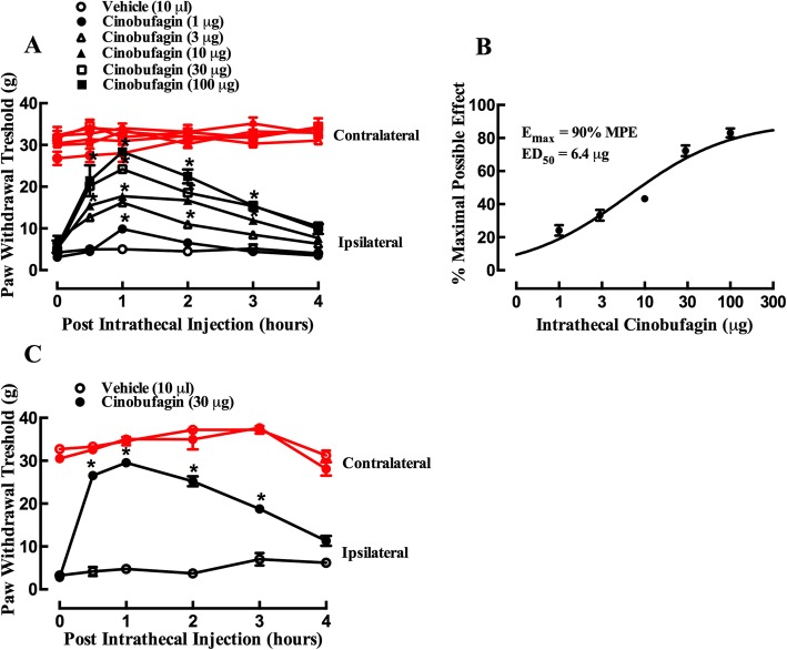
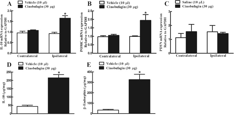
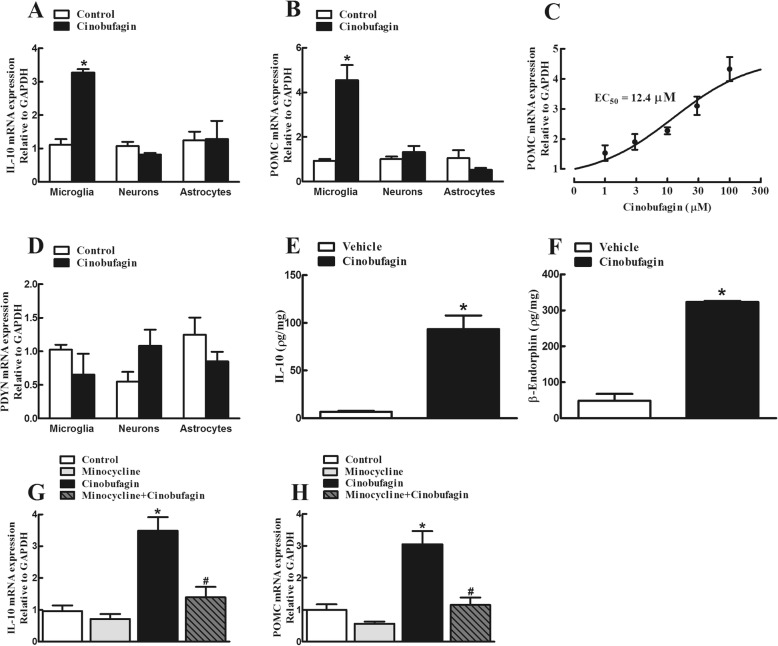
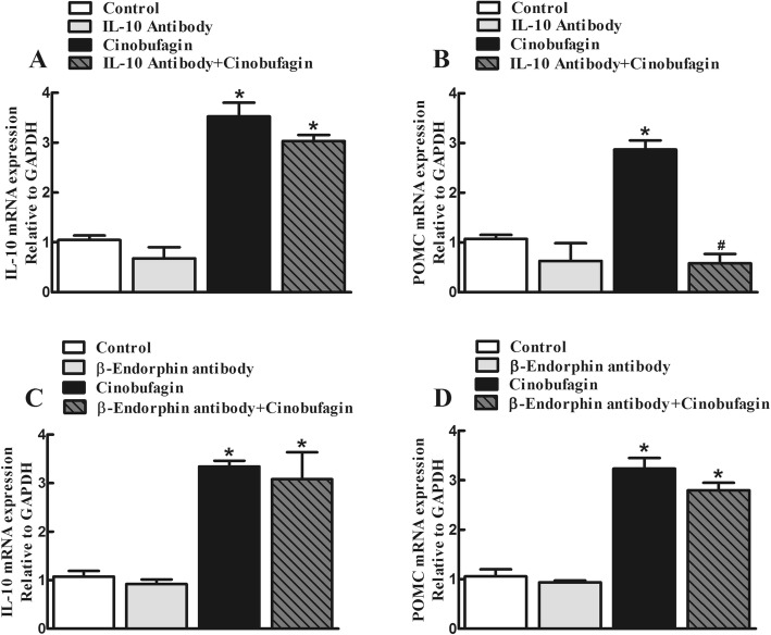
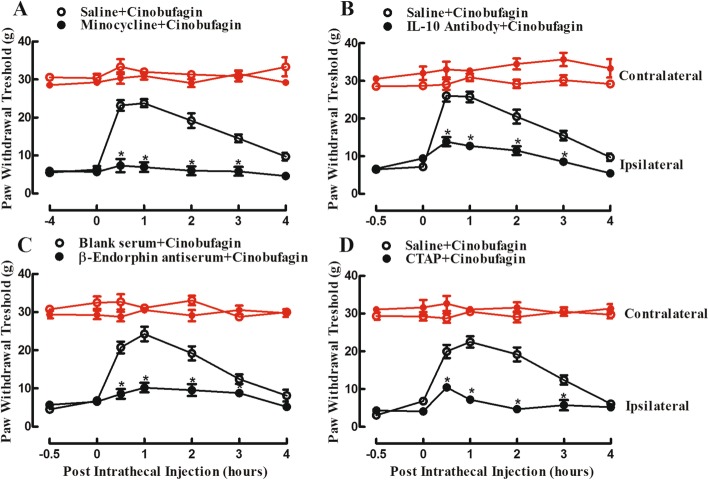
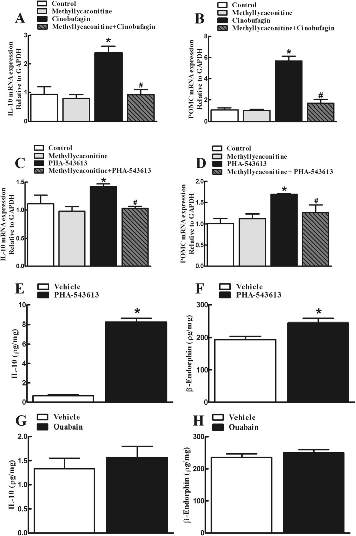
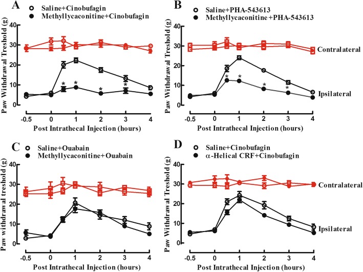
Results: Cinobufagin, given intrathecally, dose-dependently attenuated mechanical allodynia in bone cancer pain rats, with the projected Emax of 90% MPE and ED50 of 6.4 μg. Intrathecal cinobufagin also stimulated the gene and protein expression of IL-10 and β-endorphin (but not dynorphin A) in the spinal cords of bone cancer pain rats. In addition, treatment with cinobufagin in cultured primary spinal microglia but not astrocytes or neurons stimulated the mRNA and protein expression of IL-10 and β-endorphin, which was prevented by the pretreatment with the IL-10 antibody but not β-endorphin antiserum. Furthermore, spinal cinobufagin-induced mechanical antiallodynia was inhibited by the pretreatment with intrathecal injection of the microglial inhibitor minocycline, IL-10 antibody, β-endorphin antiserum and specific μ-opioid receptor antagonist CTAP. Lastly, cinobufagin- and the specific α-7 nicotinic acetylcholine receptor (α7-nAChR) agonist PHA-543613-induced microglial gene expression of IL-10/β-endorphin and mechanical antiallodynia in bone cancer pain were blocked by the pretreatment with the specific α7-nAChR antagonist methyllycaconitine.

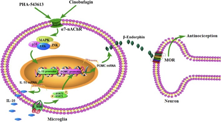
Conclusions: Our results illustrate that cinobufagin produces mechanical antiallodynia in bone cancer pain through spinal microglial expression of IL-10 and subsequent β-endorphin following activation of α7-nAChRs. Our results also highlight the broad significance of the recently uncovered spinal microglial IL-10/β-endorphin pathway in antinociception.

Keywords: Cinobufagin; IL-10/β-endorphin pathway; Microglia; α7-nicotinic acetylcholine receptor (α7-nAChR).


Conflict of interest statement

The authors declare that they have no competing interests.

Figures

Fig. 1
Fig. 1
Inhibitory effects of cinobufagin, given intrathecally, on mechanical allodynia in the rat model of bone cancer pain. The rats were inoculated with cancer cells for approximately 3 weeks and their mechanical thresholds were then measured by using electric von Frey filaments in both the contralateral and ipsilateral hindpaws. a Female bone cancer pain rats received single intrathecal administration of saline or cinobufagin (1, 3, 10, 30, or 100 μg). b Dose-response analysis of cinobufagin on mechanical allodynia in the ipsilateral hindpaws of female bone cancer pain rats 1 h after its injection, best projected by the non-linear least-squares method. c Male bone cancer pain rats received single intrathecal administration of saline or cinobufagin (30 μg). The data are presented as means ± SEM (n = 6 per group). The asterisk denotes statistical significance (p < 0.0001) compared to the saline control group, by repeated-measured two-way ANOVA followed by the post hoc Student–Newman–Keuls test
Fig. 2
Fig. 2
Effects of cinobufagin on spinal gene (ac) and protein (d, e) expression of IL-10, β-endorphin and dynorphin A in bone cancer pain rats. The spinal lumbar enlargement (L3-L5) of female bone cancer pain rats were collected 1 h after intrathecal injection of cinobufagin. The mRNA expression of IL-10, the β-endorphin precursor proopiomelanocortin (POMC) and dynorphin precursor prodynorphin (PDYN), and protein expression of IL-10 and β-endorphin were measured by using qRT-PCR and commercial fluorescent immunoassay kits, respectively. The data are presented as means ± SEM (n = 6 per group). The asterisk denotes statistical significance (p < 0.05) compared to the saline control group, by unpaired and two-tailed Student t test
Fig. 3
Fig. 3
Effects of cinobufagin on the gene (ad) and protein (e, f) expression of IL-10, the β-endorphin precursor proopiomelanocortin (POMC) and dynorphin precursor prodynorphin (PDYN) in the primary cultures of spinal microglia, neurons, and astrocytes. g, h The blockade effect of the microglial inhibitor minocycline on cinobufagin-induced gene overexpression of IL-10 and POMC. The primary culture cells, originated from the spinal cords of 1-day-old neonatal rats, were collected 2 h after cinobufagin incubation. For the blockade study, minocycline was incubated 1 h prior to cinobufagin treatment. The mRNA expression of IL-10, POMC, and PDYN was measured by using qRT-PCR. The data are presented as means ± SEM (n = 3 independent repeats with duplicates). The asterisk and number sign denote statistical significance (p < 0.001) compared to the control group and cinobufagin group, respectively, by unpaired and two-tailed Student t test (af) or one-way ANOVA followed by the post hoc Student–Newman–Keuls test (g, h)
Fig. 4
Fig. 4
Effects of the IL-10 antibody (a, b) and β-endorphin antiserum (c, d) on cinobufagin-stimulated gene expression of IL-10 and β-endorphin precursor proopiomelanocortin (POMC) in primary cultures of spinal microglia. Microglia cells, originated from the spinal cords of 1-day-old neonatal rats, were first treated with the IL-10 antibody or β-endorphin antiserum for 2 h followed by cinobufagin, and were collected 2 h after the cinobufagin treatment. The mRNA expression of IL-10 and POMC was measured using qRT-PCR. The data are presented as means ± SEM (n = 3 independent repeats with duplicates). The asterisk denotes statistical significance (p < 0.001) compared to the control group, by unpaired and two-tailed Student t test
Fig. 5
Fig. 5
Blockade effects of intrathecal injection of the microglial inhibitor minocycline (a), IL-10 antibody (b), β-endorphin antiserum (c), and selective μ-opioid receptor CTAP (d) on spinal cinobufagin-induced mechanical antiallodynia in the rat model of bone cancer pain. Female bone cancer pain rats, approximately 3 weeks after cancer cell inoculation, received two intrathecal injections, and mechanical thresholds in both the contralateral and ipsilateral hindpaws were measured by using electric von Frey filaments. Minocycline was intrathecally injected 4 h prior to cinobufagin treatment, whereas the IL-10 antibody, β-endorphin antiserum, and CTAP were intrathecally given 30 min before cinobufagin injection. Withdrawal thresholds were measured in both the contralateral and ipsilateral hindpaws. The data are presented as means ± SEM (n = 6 per group). The asterisk denotes statistical significance (p < 0.05) compared to the vehicle control group, by repeated-measured two-way ANOVA followed by the post hoc Student–Newman–Keuls test
Fig. 6
Fig. 6
Blockade effects of the specific α7-nicotinic acetylcholine receptor (α7-nAChR) antagonist methyllycaconitine on cinobufagin (a, b)- and the specific α7-nAChR agonist PHA-543613 (c, d)-stimulated gene expression of IL-10 and the β-endorphin precursor proopiomelanocortin (POMC) in primary cultures of spinal microglia. eh. Effects of PHA-543613 and ouabain on the protein expression of IL-10 and β-endorphin. Microglial cells, originated from the spinal cords of 1-day-old neonatal rats, were incubated with cinobufagin (100 μM), PHA-543613 (100 μM), or ouabain (10 μM). The mRNA and protein expression of IL-10 and POMC was measured 2 h later using qRT-PCR and the commercial kits, respectively. For the methyllycaconitine blockade study, methyllycaconitine was incubated 0.5 h prior to cinobufagin or PHA-543613 treatment. The data are presented as means ± SEM (n = 3 independent repeats with duplicates). The asterisk and number sign denote statistical significance (p < 0.001) compared to the control and cinobufagin group, respectively, by one-way ANOVA followed by the post hoc Student–Newman–Keuls test
Fig. 7
Fig. 7
Blockade effects of the specific p38 mitogen-activated protein kinase (MAPK) activation inhibitor SB203580 (a, b), JNK MAPK activation inhibitor SP600125 (c, d), and ERK1/2 MAPK activation inhibitor UO126 (e, f) on cinobufagin-stimulated gene overexpression of IL-10 and the β-endorphin precursor proopiomelanocortin (POMC) in primary cultures of spinal microglia. Cultured primary microglial cells, originated from the spinal cords of 1-day-old neonatal rats, incubated with each MAPK inhibitor for 1 hour followed by cinobufagin (100 μM) for 2 h. The mRNA expression of IL-10 and POMC was measured using qRT-PCR. The data are presented as means ± SEM (n = 3 independent repeats with duplicates). The asterisk and number sign denote statistical significance (p < 0.001) compared to the control group and cinobufagin group, respectively, by one-way ANOVA followed by the post hoc Student–Newman–Keuls test
Fig. 8
Fig. 8
Effects of the specific α7-nicotinic acetylcholine receptor (α7-nAChR) antagonist methyllycaconitine (ac) and CRF receptor antagonist α-helical CRF (9-41) (d) on cinobufagin-, PHA-543613-, or ouabain-induced mechanical antiallodynia in the rat model of bone cancer pain. Female bone cancer pain rats, approximately 3 weeks after cancer cell inoculation, received intrathecal injection of the vehicle (10 μL), methyllycaconitine (10 μg) or α-helical CRF (9-41) (20 μg) 0.5 h later followed by cinobufagin (30 μg), PHA-543613 (12 μg), or ouabain (2.5 μg), and mechanical thresholds in both the contralateral and ipsilateral hindpaws were measured by using electric von Frey filaments. The data are presented as means ± SEM (n = 6 per group). The asterisk denotes statistical significance (p < 0.05) compared to the vehicle control group, by repeated-measured two-way ANOVA followed by the post hoc Student–Newman–Keuls test
Fig. 9
Fig. 9
Illustration of the proposed mechanisms underlying cinobufagin-mediated antinociception through stimulation of spinal microglial IL-10/β-endorphin pathway following α7-nicotinic acetylcholine receptor (α7-nAChR) activation. Cinobufagin and PHA-543613 stimulate mitogen-activated protein kinase (MAPK) activation following agonism of the α7-nAChR and excrete IL-10, which subsequently activates the IL-10 receptor and expresses β-endorphin through an autocrine mechanism. The released β-endorphin passes the microglial neuronal synapse and activates μ-opioid receptor (MOR) in neurons to produce antinociception

References

    1. AlSharari SD, Freitas K, Damaj MI. Functional role of alpha7 nicotinic receptor in chronic neuropathic and inflammatory pain: studies in transgenic mice. Biochem Pharmacol. 2013;86(8):1201–1207. - PubMed
    1. Baek SH, Kim C, Lee JH, Nam D, Lee J, Lee S-G, Ahn KS. Cinobufagin exerts anti-proliferative and pro-apoptotic effects through the modulation ROS-mediated MAPKs signaling pathway. Immunopharmacol Immunotoxicol. 2015;37(3):265–273. - PubMed
    1. Berta T, Qadri Y, Chen G, Ji R. Microglial signaling in chronic pain with a special focus on caspase 6, p38 MAP kinase, and sex dependence. J Dent Res. 2016;95(10):1124–1131. - PMC - PubMed
    1. Bick RJ, Poindexter BJ, Sweney RR, Dasgupta A. Effects of Chan Su, a traditional Chinese medicine, on the calcium transients of isolated cardiomyocytes: cardiotoxicity due to more than Na, K-ATPase blocking. Life Sci. 2002;72(6):699–709. - PubMed
    1. Boscia F, Begum G, Pignataro G, Sirabella R, Cuomo O, Casamassa A, Annunziato L. Glial Na+ dependent ion transporters in pathophysiological conditions. Glia. 2016;64(10):1677–1697. - PMC - PubMed