Review
. 2020 Aug:35:101454.
doi: 10.1016/j.redox.2020.101454. Epub 2020 Feb 9.

Lactate as a fulcrum of metabolism

Affiliations
Review

Lactate as a fulcrum of metabolism

George A Brooks. Redox Biol. 2020 Aug.

Abstract

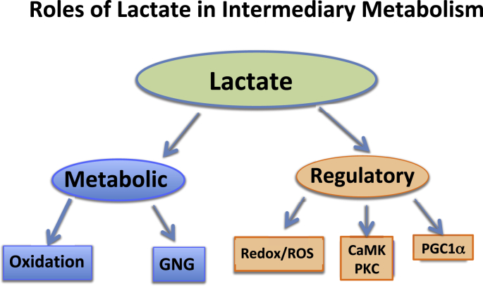
Mistakenly thought to be the consequence of oxygen lack in contracting skeletal muscle we now know that the L-enantiomer of the lactate anion is formed under fully aerobic conditions and is utilized continuously in diverse cells, tissues, organs and at the whole-body level. By shuttling between producer (driver) and consumer (recipient) cells lactate fulfills at least three purposes: 1] a major energy source for mitochondrial respiration; 2] the major gluconeogenic precursor; and 3] a signaling molecule. Working by mass action, cell redox regulation, allosteric binding, and reprogramming of chromatin by lactylation of lysine residues on histones, lactate has major influences in energy substrate partitioning. The physiological range of tissue [lactate] is 0.5-20 mM and the cellular Lactate/Pyruvate ratio (L/P) can range from 10 to >500; these changes during exercise and other stress-strain responses dwarf other metabolic signals in magnitude and span. Hence, lactate dynamics have rapid and major short- and long-term effects on cell redox and other control systems. By inhibiting lipolysis in adipose via HCAR-1, and muscle mitochondrial fatty acid uptake via malonyl-CoA and CPT1, lactate controls energy substrate partitioning. Repeated lactate exposure from regular exercise results in major effects on the expression of regulatory enzymes of glycolysis and mitochondrial respiration. Lactate is the fulcrum of metabolic regulation in vivo.

Keywords: Aerobic; Anaerobic; Cell-cell signaling; Energy substrate partitioning; Exercise; Gluconeogenesis; Glycolysis; HCAR1; Histone lactylation; Mitochondrial biogenesis; Oxidative metabolism; PGC-1α; PPAR-γ; SIRT activation; TGFβ.


Conflict of interest statement

Declaration of competing interest The author has no competing interests to declare.

Figures

Fig. 1
Fig. 1
Depiction of the Lactate Shuttle as it describes the roles of lactate in delivery of oxidative and gluconeogenic substrates as well as in cell signaling. Examples of the Cell-Cell Lactate Shuttles include lactate exchanges between producer (or driver) white-glycolytic (FW) and red-oxidative (SO) consumer (or recipient) fibers within a working muscle bed and between producer working skeletal muscle and consumer heart, brain, liver and kidneys. Examples of Intracellular Lactate Shuttles include cytosol-mitochondrial and cytosol-peroxisome exchanges. Indeed most, if not all, lactate shuttles are driven by a concentration or pH gradient, or by redox state. Symbols: G – Glucose and Glycogen, L – Lactate, and M – elements of the mitochondrial reticulum. Originally compiled from diverse sources (16, 18, 19); from (22).
Fig. 2
Fig. 2
Depiction of the Lactate Shuttle as it fulfills three physiological Functions: 1] lactate is a major energy source; 2] lactate is the major gluconeogenic precursor; and 3] lactate is a signaling molecule with autocrine-, paracrine- and endocrine-like effects and has been called a “lactormone”. These functions can be subdivided to Metabolic (oxidative fuel) and gluconeogenic (GNG) functions, and Regulatory, or signaling functions.
Fig. 3
Fig. 3
There is strong evidence that glucose and glycogen catabolism proceed to lactate production under fully aerobic conditions in studies on intact animals, animal tissue preparations and healthy humans in vivo. In muscles and arterial blood of resting healthy humans lactate concentration approximates 1.0 mM, while pyruvate concentration approximates 0.1 mM, the Lactate/Pyruvate (L/P) being 10, with net lactate production and release from resting muscle of healthy individuals when intramuscular partial pressure of oxygen (PO2) approximates 40 Torr, well above the critical mitochondrial PO2 for maximal mitochondrial respiration (1–2 Torr) [7,39,140]. During exercise at about 65% of maximal oxygen consumption (VO2max) lactate production and net lactate release from working muscle beds rise and the L/P rises more than an order of magnitude (to ~500), but the intramuscular PO2 remains at 3–4 Torr, well above the critical mitochondrial O2 level. Hence, it is appropriate to conclude that in healthy humans glycolysis proceeds to lactate under fully aerobic conditions. Importantly, most (75–80%) of lactate is disposed of immediately within the tissue or subsequent to release and reuptake by working muscle, with significant uptake and oxidation by heart or oxidation and liver for gluconeogenesis. Adapted from diverse sources [14,39,80,136,159]; from Ref. [22].
Fig. 4
Fig. 4
A schematic showing the putative mitochondrial lactate oxidation complex (mLOC): MCT1 is inserted into the mitochondrial inner membrane strongly interacting with its chaperone protein CD147, and is also associated with COX as well as mitochondrial LDH (mLDH) which could be located at the outer side of the inner membrane. Lactate, which is always produced in cytosol of muscle and other tissues because of the abundance, activity, and characteristics of cytosolic LDH, is oxidized to pyruvate via the lactate oxidation complex in mitochondria of the same cell. This endergonic lactate oxidation reaction is coupled to the exergonic redox change in COX during mitochondrial electron transport. Abbreviations: GP, glycerol phosphate; Mal-Asp, malate-aspartate; MCT, monocarboxylate (lactate) transporter, mPC, mitochondrial pyruvate carrier, ETC, electron transport chain; TCA, tricarboxylic acid; adapted from Ref. [73].
Fig. 5
Fig. 5
Schematic summarizing the effects of lactate on intracellular signaling in muscle. Contractions stimulate glycolysis and subsequent lactate production and accumulation. In combination, lactate accumulation and mitochondrial respiration induce ROS production. A ROS-sensitive transcriptome is activated, which elicits many cell responses seen in the response to exercise, including MCT1 expression, mitochondrial biogenesis, and the production of antioxidant enzymes (e.g., GPx). In this figure, novel signaling effects described in the present report (solid arrows) are shown. ROS generation (left side of figure) is responsible for regulating MCT1 expression. For mitochondrial biogenesis (right side), it is likely that the lactate-signaling pathway merges with calcium (Ca++) signaling as contractions increase cytosolic Ca++ flux. By itself, lactate increases expressions of slow type troponin I (TnI) and myogenin that are also known to be responsive to Ca++ flux via calcineurin (CaN). ROS can increase intracellular Ca++ that raises CaMK activity. As well, free Ca++ can also activate CaMK. Lactate elicits a large number of adaptive responses, which coordinate metabolism as a functional adaptation to exercise in skeletal muscle cells such as proliferation of the lactate oxidation complex; from Ref. [75].
Fig. 6
Fig. 6
Illustration of how lactatemia affects blood [glucose] and peripheral glucose uptake as well as the production, uptake and oxidation of FFA giving rise to metabolic inflexibility in muscle. Lactate is the inevitable consequence of glycolysis [139], the minimal muscle L/P being 10 and rising to an L/P > 100 when glycolytic flux is high [80]. Lactate is the favored oxidizable substrate, provides product inhibition of glucose and FFA oxidation. As the products of glycolysis, lactate and pyruvate provide negative feedback inhibition of glucose disposal (blue dashed lines). Also as the predominant mitochondrial substrate, lactate gives rise to Acetyl-CoA, and in turn Malonyl-CoA. Acetyl-CoA inhibits β-ketothiolase, and hence β-oxidation, while Malonyl-CoA inhibits mitochondrial FFA-derivative uptake via CPT1 (T) [142]. Moreover, lactate is the main gluconeogenic precursor raising glucose production and blood [glucose] (red lines). Via GPR81 binding, lactate inhibits lipolysis in WAT (T) depressing circulating [FFA] [84,99]. This model explains the paradoxical presence of lactatemia in high intensity exercise and insulin resistant states with limited ability to oxidize fat (green lines). Modified from Hashimoto et al. [74]. Abbreviations: CPT1-Carnitine Palmitoyl Transporter-1, FFA-Free Fatty Acid, FAT-Fatty Acid Translocator comprised of CD36 and FABPc, GLUT-Glucose Transporter, s-sarcolemmal, m-mitochondrial, Malonyl-CoA formed from exported TCA citrate controlled by the interactions of Malonyl-CoA Decarboxylase (MCD) and Acetyl-CoA Carboxylase (ACC), MCT-Monocarboxylate Transporter, mPC-Mitochondrial Pyruvate Transporter, PDH-Pyruvate Dehydrogenase, WAT-White Adipose Tissue, T-Inhibition. Not shown is Fatty Acyl-Co (FA-CoA) that will accumulate if FFAs are taken up by myocytes, but blocked from mitochondrial entry by the effect of Malonyl-CoA on CPT1. Accumulated intracellular FA-CoA will give rise to Intramyocellular Triglyceride (IMTG) and the formulation of LC-FA, DAG and Ceramides via inhibition of PI3 Kinase (PI3-k) and reducing GLUT4 translocation; from Ref. [22]. (For interpretation of the references to colour in this figure legend, the reader is referred to the Web version of this article.)
Fig. 7
Fig. 7
Lactate shuttling gone amiss. In cancer high glucose consumption leading to lactate production under fully aerobic conditions (i.e., the Warburg Effect) are features of cancer cells and tumors. Lactagenesis in cancer can be viewed as a highly orchestrated effort from oncogenes and tumor suppressor mutations for continuous and non-stop glucose utilization to produce lactate involving 5 major steps [1]: Increased glucose uptake through increased expression and translocation of glucose transporters GLUT by transcription factors Hypoxia-Inducible Factor 1α (HIF-1) and c-Myc oncogene as well as loss of expression of tumor suppression factor p53 [2]. Increased glycolytic enzyme expression and activity, especially Lactate Dehydrogenase A (LDHA) by HIF-1α, c-MYC and p53 dysregulation. 3) Decreased mitochondrial function mainly by p53 dysregulation [4]. Increased lactate production, accumulation and release due to mass effect of accelerated glycolysis, mitochondrial dysfunction and increased LDHA expression. 5) Upregulation of monocarboxylate transporters MTC1 and MCT4 and their plasma membrane chaperon, CD147, contributing to dysregulated lactate shuttling in support of carcinogenesis; from Ref. [144].

References

    1. Ahmed K., Tunaru S., Tang C., Muller M., Gille A., Sassmann A., Hanson J., Offermanns S. An autocrine lactate loop mediates insulin-dependent inhibition of lipolysis through GPR81. Cell Metabol. 2010;11:311–319. - PubMed
    1. Azevedo J.L., Tietz E., Two-Feathers T., Paull J., Chapman K. Lactate, fructose and glucose oxidation profiles in sports drinks and the effect on exercise performance. PloS One. 2007;2:e927. - PMC - PubMed
    1. Baba N., Sharma H.M. Histochemistry of lactic dehydrogenase in heart and pectoralis muscles of rat. J. Cell Biol. 1971;51:621–635. - PMC - PubMed
    1. Barros L.F. Metabolic signaling by lactate in the brain. Trends Neurosci. 2013;36:396–404. - PubMed
    1. Bayod S., Del Valle J., Lalanza J.F., Sanchez-Roige S., de Luxan-Delgado B., Coto-Montes A., Canudas A.M., Camins A., Escorihuela R.M., Pallas M. Long-term physical exercise induces changes in sirtuin 1 pathway and oxidative parameters in adult rat tissues. Exp. Gerontol. 2012;47:925–935. - PubMed

Publication types