

Pulmonary Pathology of Early-Phase 2019 Novel Coronavirus (COVID-19) Pneumonia in Two Patients With Lung Cancer
- PMID: 32114094
- PMCID: PMC7128866
- DOI: 10.1016/j.jtho.2020.02.010
Pulmonary Pathology of Early-Phase 2019 Novel Coronavirus (COVID-19) Pneumonia in Two Patients With Lung Cancer
Abstract
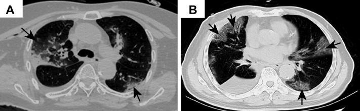
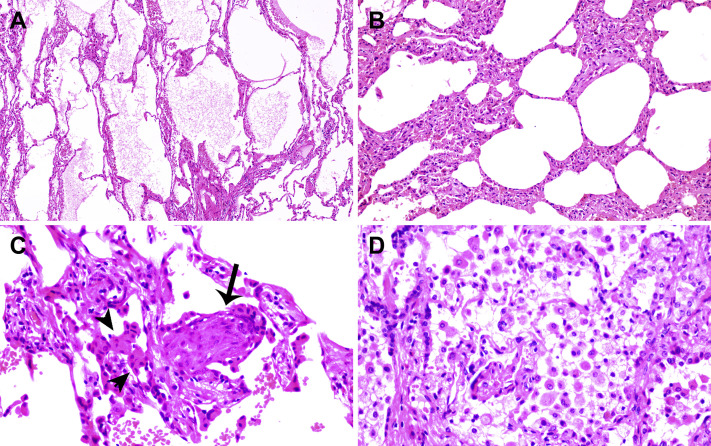
There is currently a lack of pathologic data on the novel coronavirus (severe acute respiratory syndrome coronavirus 2) pneumonia, or coronavirus disease 2019 (COVID-19), from autopsy or biopsy. Two patients who recently underwent lung lobectomies for adenocarcinoma were retrospectively found to have had COVID-19 at the time of the operation. These two cases thus provide important first opportunities to study the pathology of COVID-19. Pathologic examinations revealed that apart from the tumors, the lungs of both patients exhibited edema, proteinaceous exudate, focal reactive hyperplasia of pneumocytes with patchy inflammatory cellular infiltration, and multinucleated giant cells. Hyaline membranes were not prominent. Because both patients did not exhibit symptoms of pneumonia at the time of operation, these changes likely represent an early phase of the lung pathology of COVID-19 pneumonia.
Keywords: COVID-19 pneumonia; Coronavirus; Pathology; SARS-CoV-2.
Copyright © 2020 International Association for the Study of Lung Cancer. Published by Elsevier Inc. All rights reserved.
Figures

Comment in
-
Coronaviruses: Facts, Myths, and Hypotheses.J Thorac Oncol. 2020 May;15(5):675-678. doi: 10.1016/j.jtho.2020.02.024. Epub 2020 Mar 7. J Thorac Oncol. 2020. PMID: 32151778 Free PMC article. No abstract available.
-
Pulmonary Pathology of Early Phase 2019 Novel Coronavirus Pneumonia.J Thorac Oncol. 2020 May;15(5):e67. doi: 10.1016/j.jtho.2020.03.013. J Thorac Oncol. 2020. PMID: 32340677 Free PMC article. No abstract available.
-
Pathology of 2019 Novel Coronavirus Pneumonia: A Dynamic Disease Process.J Thorac Oncol. 2020 May;15(5):e67-e68. doi: 10.1016/j.jtho.2020.03.015. J Thorac Oncol. 2020. PMID: 32340678 Free PMC article. No abstract available.
-
Coronavirus Disease 2019 or Lung Cancer: A Differential Diagnostic Experience and Management Model From Wuhan.J Thorac Oncol. 2020 Aug;15(8):e141-e142. doi: 10.1016/j.jtho.2020.04.030. Epub 2020 May 7. J Thorac Oncol. 2020. PMID: 32387713 Free PMC article. No abstract available.
References
Publication types
MeSH terms
LinkOut - more resources
Full Text Sources
Other Literature Sources
Medical
Miscellaneous
