Review
. 2020 Apr:63:174-185.
doi: 10.1016/j.ceb.2020.01.016. Epub 2020 Feb 27.

Receptor tyrosine kinase activation: From the ligand perspective

Affiliations
Review

Receptor tyrosine kinase activation: From the ligand perspective

Raphael Trenker et al. Curr Opin Cell Biol. 2020 Apr.

Abstract

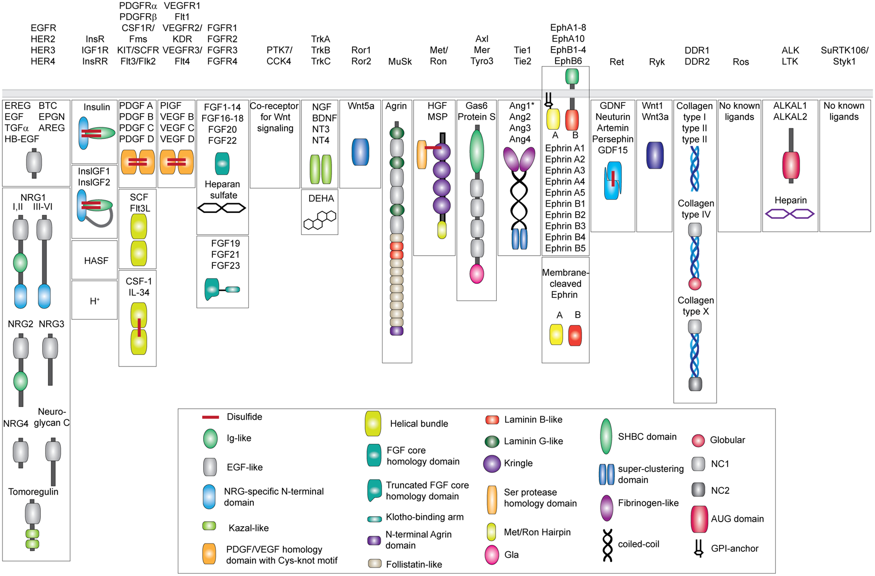
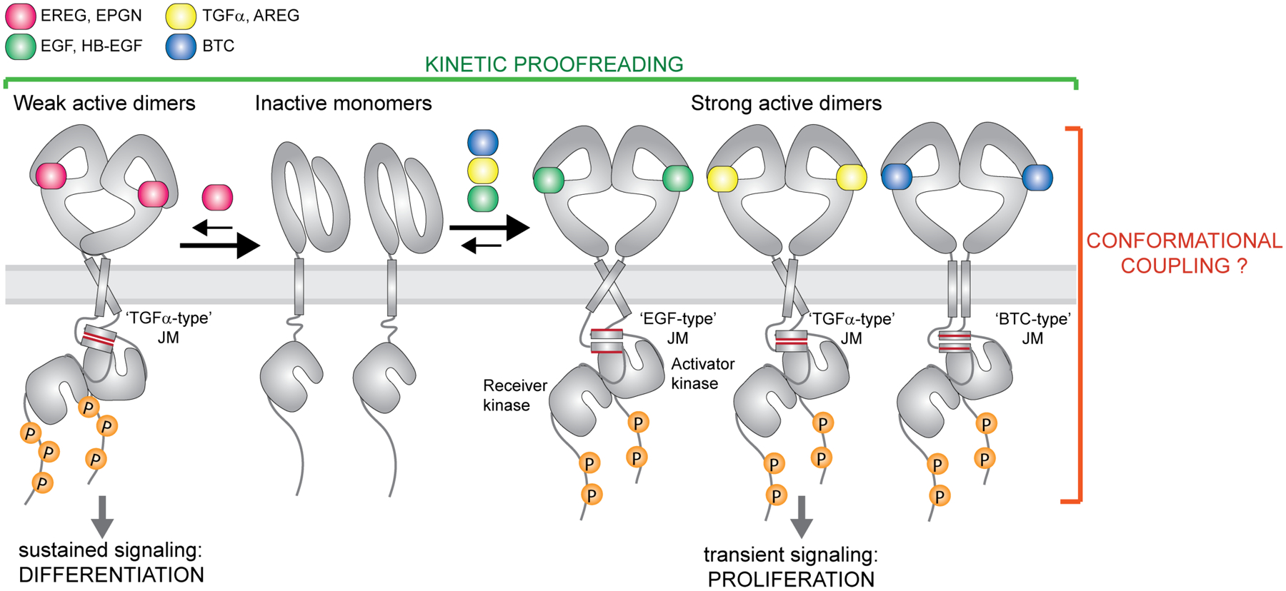
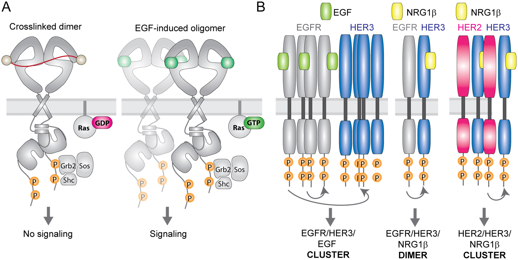
Receptor tyrosine kinases (RTKs) are single-span transmembrane receptors in which relatively conserved intracellular kinase domains are coupled to divergent extracellular modules. The extracellular domains initiate receptor signaling upon binding to either soluble or membrane-embedded ligands. The diversity of extracellular domain structures allows for coupling of many unique signaling inputs to intracellular tyrosine phosphorylation. The combinatorial power of this receptor system is further increased by the fact that multiple ligands can typically interact with the same receptor. Such ligands often act as biased agonists and initiate distinct signaling responses via activation of the same receptor. Mechanisms behind such biased agonism are largely unknown for RTKs, especially at the level of receptor-ligand complex structure. Using recent progress in understanding the structures of active RTK signaling units, we discuss selected mechanisms by which ligands couple receptor activation to distinct signaling outputs.

Keywords: Biased agonism; Growth factor; Ligand; Receptor tyrosine kinase; Signaling.


Conflict of interest statement

Conflict of interest statement Nothing declared.

Figures

Figure 1:
Figure 1:. Diversity of the human RTK ligands.
RTKs are shown on the top categorized into 19 different families as originally described [1] and recently revised to remove the LMR1–3 family due to its re-classification as Ser/Thr receptor kinases [127]. Ligands for each RTK family are shown underneath in mature, secreted form. All membrane-tethered ligands are cleaved off the membrane except for ephrins, which activate their cognate receptors in a juxtacrine fashion. The ligands are drawn with their N-terminus pointing away from the membrane. Main structural domains are depicted in a cartoon form in their known oligomeric state except for Angiotensins*, which may form higher-order oligomers in addition to dimers. If applicable, domain labels are included as captions. Sizes of individual domains are not drawn up to scale.
Figure 2:
Figure 2:. Ligand-specific signaling through EGFR homodimers.
EGFR forms high-affinity complexes with its cognate ligands Transforming Growth Factor α (TGFα), Epidermal Growth Factor (EGF), Amphiregulin (AREG), Betacellulin (BTC) and Heparin-Binding-EGF (HB-EGF) but lower-affinity complexes with Epiregulin (EREG) and Epigen (EPGN). Available structures of EGF, TGFα±EPGN and EREG bound to EGFR ECDs are so far consistent with the EGFR ECDs adopting symmetric dimers with high affinity ligands and asymmetric structures with lower affinity ligands. Weaker EGFR dimers lead to sustained signaling and cell differentiation while formation of strong complexes causes transient receptor signaling and cell proliferation. Thus, the dynamics of ligand-dependent receptor association and dissociation has an important impact on the recruitment of downstream effectors and the ligand-specific cellular responses (kinetic proofreading). Biochemical studies on the EGFR juxtamembrane (JM) segment and studies of full-length EGFR in cells are consistent with the presence of the JM coiled-coil dimer in active receptor complex. Conformation of the JM dimer appears to change depending on a bound ligand. At least three specific JM dimer modes have been described for EGFR, denoted here as ‘TGFα-type’, the ‘EGF-type’ and the ‘BTC-type’.
Figure 3:
Figure 3:. Ligands can diversify RTK signaling via promotion of higher-order receptor oligomers.
(A) Cartoon representation of active EGFR dimers in the plasma membrane dimerized via DNA-bases crosslinkers (left) or upon EGF binding (right). Both receptors undergo autophosphorylation but only EGF-bound receptors further progress into clusters and induce signaling via Ras. (B) Simplified illustration of EGFR, HER2 and HER3 oligomerization patterns upon EGF or NRG1β stimulation in cells. EGF treatment of cells co-expressing EGFR and HER3 causes phosphorylation and clustering of both receptors with HER3 phosphorylation occuring via a non-canonical mechanism that does not rely on asymmetric dimerization of the kinase domains. In contrast, NRG1β treatment induces HER3 phosphorylation by EGFR via canonical asymmetric kinase dimerization, without promoting clustering of the receptors. In yet another scenario, NRG1β stimulation of HER2/HER3 expressing cells induces clusters of both receptors in which HER3 phosphorylation is dependent on formation of asymmetric kinase dimers.
Figure 4:
Figure 4:. Co-receptors assure ligand specificity and control of downstream signaling in the FGF and RET signaling systems.
(A) Domain architecture of the 2/2/2 RET receptor/co-receptor/ligand complex. The receptor with its structural domains is shown in grey. The co-receptors GFRAL and GFRα1–4 are shown in blue and the ligands GDF15, GDNF, NRTN, ARTN, Persephin are shown in cyan. (B) Cryo-EM structures of the 2:2:2 RET ECD receptor/co-receptor/ligand complexes are shown as top and front views (PDB codes from left to right: 6Q2N, 6Q2O, 6Q2S, 6Q2J). Adapted from [113]. (C) Domain architecture of the autocrine/paracrine and endocrine active 2:2 and 2:2:2 complexes of receptor/ligand and receptor/ligand/Klotho complexes, respectively. Both representations illustrate heparan sulfate (HS) bound to the complexes. (D) Simplified overview of receptor specificity for autocrine/paracrine and endocrine ligands. Paracrine/autocrine ligands bind to HS with high affinity, which traps the ligands close to the site of secretion and enables them to bind the cognate receptors. Autocrine/paracrine ligands can be either specific towards FGFR1–3b or c isoforms or bind both isoforms promiscuously. The presence of HS is mandatory for activation by the paracrine/autocrine ligands. Endocrine ligands FGF19, FGF21 and FGF23 have low affinity for both FGFR isoforms and HS and require the presence of either α-Klotho or β-Klotho co-receptors for binding and activation of their cognate FGF receptors. The low affinity of endocrine ligands to HS facilitates secretion from the tissue of origin to distal organs that express the respective Klotho/FGFRc complexes. Despite low affinity in the absence of Klotho, HS appears to be required for receptor dimerization upon ligand binding. (E) Crystal structure of the FGFR1c/α-Klotho/FGF23 ECD complex (PDB code: 5W21). The structure illustrates tight interactions between FGF23 and the α-Klotho ECD via the Klotho binding arm, and with the receptor ECD via a truncated FGF core homology domain.

References

    1. Lemmon MA, Schlessinger J: Cell signaling by receptor tyrosine kinases. Cell 2010, 141:1117–1134. - PMC - PubMed
    1. Falls D: Neuregulins: functions, forms, and signaling strategies. Experimental Cell Research 2003, 284:14–30. - PubMed
    1. Wieduwilt MJ, Moasser MM: The epidermal growth factor receptor family: Biology driving targeted therapeutics. Cellular and Molecular Life Sciences 2008, 65:1566–1584. - PMC - PubMed
    1. Flanagan JG, Gale NW, Hunter T, Pasquale EB, TessierLavigne M: Unified nomenclature for Eph family receptors and their ligands, the ephrins. Cell 1997, 90:403–404. - PubMed
    1. Pasquale EB: Eph receptor signalling casts a wide net on cell behaviour. Nature Reviews Molecular Cell Biology 2005, 6:462–475. - PubMed

Publication types

MeSH terms