Review
. 2020 Mar;13(3):100738.
doi: 10.1016/j.tranon.2019.12.010. Epub 2020 Feb 27.

Resistance to Checkpoint Inhibition in Cancer Immunotherapy

Affiliations
Review

Resistance to Checkpoint Inhibition in Cancer Immunotherapy

Luisa Barrueto et al. Transl Oncol. 2020 Mar.

Abstract

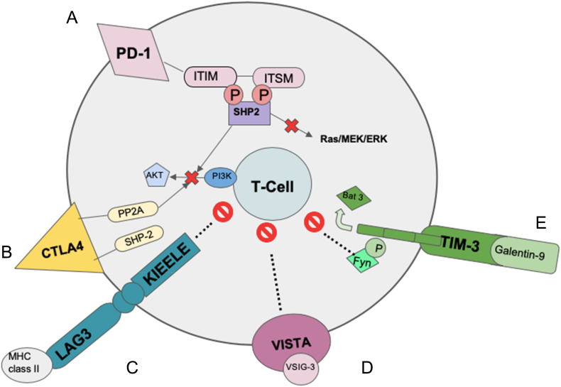
The interaction of the host immune system with tumor cells in the tissue microenvironment is essential in understanding tumor immunity and development of successful cancer immunotherapy. The presence of lymphocytes in tumors is highly correlated with an improved outcome. T cells have a set of cell surface receptors termed immune checkpoints that when activated suppress T cell function. Upregulation of immune checkpoint receptors such as programmed cell death 1 (PD-1) and cytotoxic T lymphocyte associated protein 4 (CTLA-4) occurs during T cell activation in an effort to prevent damage from an excessive immune response. Immune checkpoint inhibitors allow the adaptive immune system to respond to tumors more effectively. There has been clinical success in different types of cancer blocking immune checkpoint receptors such as PD-1 and CTLA. However, relapse has occurred. The innate and acquired/therapy induced resistance to treatment has been encountered. Aberrant cellular signal transduction is a major contributing factor to resistance to immunotherapy. Combination therapies with other co-inhibitory immune checkpoints such as TIM-3, LAG3 and VISTA are currently being tested to overcome resistance to cancer immunotherapy. Expression of TIM-3 has been associated with resistance to PD-1 blockade and combined blockade of TIM-3 and PD-1 has demonstrated improved responses in preclinical models. LAG3 blockade has the potential to increase the responsiveness of cytotoxic T-cells to tumors. Furthermore, tumors that were found to express VISTA had an increased rate of growth due to the T cell suppression. The growing understanding of the inhibitory immune checkpoints' ligand biology, signaling mechanisms, and T-cell suppression in the tumor microenvironment continues to fuel preclinical and clinical advancements in design, testing, and approval of agents that block checkpoint molecules. Our review seeks to bridge fundamental regulatory mechanisms across inhibitory immune checkpoint receptors that are of great importance in resistance to cancer immunotherapy. We will summarize the biology of different checkpoint molecules, highlight the effect of individual checkpoint inhibition as anti-tumor therapies, and outline the literatures that explore mechanisms of resistance to individual checkpoint inhibition pathways.


Figures

Figure 1
Figure 1
Schematic representation of immune checkpoints and their signaling in T cells A. PD-1: PD-1 ligation leads to tyrosine phosphorylation of the cytoplasmic tail of PD-1. This recruits SHP-2 to the cytoplasmic tail of PD-1. Activation of SHP-2 leads to dephosphorylation of TCR proximal kinases, causing inhibition of downstream signaling pathways , . B. CTLA4: CTLA4 binds to B7 with a higher affinity than CD28, it then interacts with the tyrosine phosphatase SHP-2 and the serine/threonine phosphatase PP2A to inhibit T cells , . C. LAG3: LAG3 binds MHC class II with greater affinity than does CD4. The signaling pathway for LAG3 has not been identified but the KIEELE motif has been shown to be essential for its inhibitory effects on CD4 T cells. Neither its effect on Tregs nor the intracellular proteins that bind it have yet to be identified . D. VISTA: Research has shown that VISTA can function as both a receptor and a ligand, but another ligand, VSIG-3 has also been discovered. Once bound to VSIG-3, VISTA is able to inhibit T cell proliferation and cytokine production . E. TIM-3: The binding of TIM-3 to its ligand Galectin-9 from tumor cells leads to phosphorylation of TIM 3 and the release of Bat-3. The release of Bat-3 allows for the interaction of TIM-3 with Fyn thereby mediating Th1 cell anergy . Abbreviations: Bat3 = HLA (Human Leukocyte Antigen) B-associated transcript 3; CD = cluster of differentiation; CTLA-4 = cytotoxic T-lymphocyte-associated protein 4; ITIM = immunoreceptor tyrosine-based inhibitory motif; ITSM = immunoreceptor tyrosine-based switch motif; LAG3 = lymphocyte-activation gene 3; MHC = major histocompatibility class; PI3K = phosphoinositide 3-kinase; P = phosphate; PD-1 = programmed cell death ligand-1; PP2A = protein phosphatase 2; SHP2 = Src homology phosphatase 2; TCR = T cell receptor; Th1 = Type 1 helper T cell; TIM-3= T cell immunoglobulin and mucin domain-3; Tregs = regulatory T cells; VISTA = V domain-containing immunoglobulin suppressor of T-cell activation; VSIG-3 = V-set and immunoglobulin domain containing 3.

References

    1. Gatti-Mays ME, Redman JM, Collins JM, Bilusic M. Cancer vaccines: Enhanced immunogenic modulation through therapeutic combinations. Hum Vaccin Immunother. 2017;13:2561–2574. - PMC - PubMed
    1. Rogers LM, Veeramani S, Weiner GJ. Complement in monoclonal antibody therapy of cancer. Immunol Res. 2014;59(0):203-210. - PMC - PubMed
    1. Showalter A, Limaye A, Oyer JL, Igarashi R, Kittipatarin C, Copik AJ, Khaled AR. Cytokines in immunogenic cell death: Applications for cancer immunotherapy. Cytokine. 2017;97:123–132. - PMC - PubMed
    1. Yang JC, Rosenberg SA. Adoptive T-cell therapy for cancer. Adv Immunol. 2016;130:279–294. - PMC - PubMed
    1. De Sousa Linhares A, Leitner J, Grabmeier-Pfistershammer K, Steinberger P. Not all immune checkpoints are created equal. Front Immunol. 2018;9:1909. - PMC - PubMed