. 2020 Mar 3;9(5):e014628.
doi: 10.1161/JAHA.119.014628. Epub 2020 Mar 2.

Arrhythmogenic Right Ventricular Cardiomyopathy: Characterization of Left Ventricular Phenotype and Differential Diagnosis With Dilated Cardiomyopathy

Affiliations

Arrhythmogenic Right Ventricular Cardiomyopathy: Characterization of Left Ventricular Phenotype and Differential Diagnosis With Dilated Cardiomyopathy

Alberto Cipriani et al. J Am Heart Assoc. .

Abstract

Background This study assessed the prevalence of left ventricular (LV) involvement and characterized the clinical, electrocardiographic, and imaging features of LV phenotype in patients with arrhythmogenic right ventricular cardiomyopathy (ARVC). Differential diagnosis between ARVC-LV phenotype and dilated cardiomyopathy (DCM) was evaluated. Methods and Results The study population included 87 ARVC patients (median age 34 years) and 153 DCM patients (median age 51 years). All underwent cardiac magnetic resonance with quantitative tissue characterization. Fifty-eight ARVC patients (67%) had LV involvement, with both LV systolic dysfunction and LV late gadolinium enhancement (LGE) in 41/58 (71%) and LV-LGE in isolation in 17 (29%). Compared with DCM, the ARVC-LV phenotype was statistically significantly more often characterized by low QRS voltages in limb leads, T-wave inversion in the inferolateral leads and major ventricular arrhythmias. LV-LGE was found in all ARVC patients with LV systolic dysfunction and in 69/153 (45%) of DCM patients. Patients with ARVC and LV systolic dysfunction had a greater amount of LV-LGE (25% versus 13% of LV mass; P<0.01), mostly localized in the subepicardial LV wall layers. An LV-LGE ≥20% had a 100% specificity for diagnosis of ARVC-LV phenotype. An inverse correlation between LV ejection fraction and LV-LGE extent was found in the ARVC-LV phenotype (r=-0.63; P<0.01), but not in DCM (r=-0.01; P=0.94). Conclusions LV involvement in ARVC is common and characterized by clinical and cardiac magnetic resonance features which differ from those seen in DCM. The most distinctive feature of ARVC-LV phenotype is the large amount of LV-LGE/fibrosis, which impacts directly and negatively on the LV systolic function.

Keywords: arrhythmogenic right ventricular cardiomyopathy; cardiac magnetic resonance; dilated cardiomyopathy; late gadolinium enhancement; left ventricular involvement; left ventricular phenotype.


Figures

Figure 1
Figure 1
Classification of the study patients according to LV systolic dysfunction and LV LGE. An overlapping LV phenotype was found in 47% of ARVC patients and 45% of DCM patients who showed both LV systolic dysfunction and LV LGE. ARVC indicates arrhythmogenic right ventricular cardiomyopathy; DCM, dilated cardiomyopathy; LGE, late gadolinium enhancement; LV, left ventricular.
Figure 2
Figure 2
Amount of LGE in ARVC vs DCM. Box plot showing difference of the amount of LV LGE between ARVC‐ and DCM‐LV phenotype (24.6% vs 13.1%, P<0.01) (A). ROC curve showing the ability of LGE percentage in distinguishing the ARVC‐ from DCM‐LV phenotype: the AUC was 0.84 (SE=0.05, 95% CI 0.75–0.93, P<0.01). The best cut‐off value of LGE percentage was 20%, which provided a sensitivity of 68% and a specificity of 100% for diagnosis of ARVC‐LV phenotype (B). ARVC indicates arrhythmogenic right ventricular cardiomyopathy; AUC, area under the curve; DCM, dilated cardiomyopathy; LGE, late gadolinium enhancement; LV, left ventricular; ROC, receiver operating characteristic.
Figure 3
Figure 3
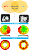
Left ventricular regional and wall layer distribution of LGE in ARVC‐ and DCM‐LV phenotype. Comparison between CMR findings in the subgroup of ARVC (left) and DCM (right) patients with overlapping LV phenotypes (ie, combination of LV systolic dysfunction and LV LGE). Patients with ARVC‐LV phenotype had lower LV volume and mass, less depressed LVEF (not shown) and greater amount of LV LGE (ie, “hypokinetic, non‐dilated and fibrotic left ventricle”) compared with DCM patients. LV LGE predominately affected the infero‐lateral segments in ARVC‐LV phenotype and septal segments in DCM. Compared with DCM, in ARVC the LV LGE significantly more often appeared as a stria pattern than a spot/patchy pattern and more frequently affected the subepicardial layers. ARVC indicates arrhythmogenic right ventricular cardiomyopathy; CMR, cardiac magnetic resonance; DCM, dilated cardiomyopathy; LGE, late gadolinium enhancement; LV, left ventricular; LVEF, left ventricular ejection fraction.
Figure 4
Figure 4
Relationship between the amount of LV LGE and LVEF. Graphs (regression plot—left, joint plot—right) showing the relationship between the amount of LV LGE and LVEF in ARVC‐ (A) and DCM‐LV phenotype (B). LGE indicates late gadolinium enhancement; LV, left ventricular; LVEF, left ventricular ejection fraction.
Figure 5
Figure 5
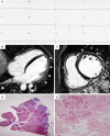
Electrocardiographic, CMR imaging, and histological features of a representative patient with ARVC undergoing cardiac transplantation. Basal ECG showing low voltages in limb leads and flattened T‐waves in the inferolateral leads (A). Post‐contrast CMR images in long‐axis (B) and short‐axis (C) views showing normal LV cavity size and subepicardial LGE (white arrows) involving the LV free wall (boxed area) and septum, from basal to apical regions (white arrows). Histologic examination of the boxed area showing fibro‐fatty myocardial replacement affecting the subepicardial LV layer (Heidenhain trichrome stain) (D); close up detailing residual myocytes embedded within fibrous and fatty tissue (E.E stain) (E). ARVC indicates arrhythmogenic right ventricular cardiomyopathy; CMR, cardiac magnetic resonance; LGE, late gadolinium enhancement; LV, left ventricular.
Figure 6
Figure 6
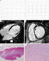
Electrocardiographic, CMR imaging, and histological features of a representative patient with DCM undergoing cardiac transplantation. Basal ECG showing complete left bundle branch block (A). Post‐contrast CMR images in long‐axis (B) and short‐axis (C) view showing a severe dilatation LV cavity and myocardial LGE (black arrows), confined to the anteroseptal region (boxed area). Histologic examination of the boxed area confirming a patchy midmural fibrosis (Heidenhain trichrome stain) (D); close up showing dilated myocytes (normal cell count) with dysmetric and dysmorphic nuclei (E.E stain) (E). CMR indicates cardiac magnetic resonance; DCM, dilated cardiomyopathy; LGE, late gadolinium enhancement; LV, left ventricular.

References

    1. Marcus FI, Fontaine GH, Guiraudon G, Frank R, Laurenceau JL, Malergue C, Grosgogeat Y. Right ventricular dysplasia: a report of 24 adult cases. Circulation. 1982;65:384–398. - PubMed
    1. Thiene G, Nava A, Corrado D, Rossi L, Pennelli N. Right ventricular cardiomyopathy and sudden death in young people. N Engl J Med. 1988;318:129–133. - PubMed
    1. Basso C, Thiene G, Corrado D, Angelini A, Nava A, Valente M. Arrhythmogenic right ventricular cardiomyopathy. Dysplasia, dystrophy, or myocarditis? Circulation. 1996;94:983–991. - PubMed
    1. Corrado D, Basso C, Thiene G, McKenna WJ, Davies MJ, Fontaliran F, Nava A, Silvestri F, Blomstrom‐Lundqvist C, Wlodarska EK, Fontaine G, Camerini F. Spectrum of clinicopathologic manifestations of arrhythmogenic right ventricular cardiomyopathy/dysplasia: a multicenter study. J Am Coll Cardiol. 1997;30:1512–1520. - PubMed
    1. Sen‐Chowdhry S, Syrris P, Prasad SK, Hughes SE, Merrifield R, Ward D, Pennell DJ, McKenna WJ. Left‐dominant arrhythmogenic cardiomyopathy: an under‐recognized clinical entity. J Am Coll Cardiol. 2008;52:2175–2187. - PubMed

Publication types

LinkOut - more resources