

An open-source framework for pulmonary fissure completeness assessment
- PMID: 32115275
- PMCID: PMC7363554
- DOI: 10.1016/j.compmedimag.2020.101712
An open-source framework for pulmonary fissure completeness assessment
Abstract
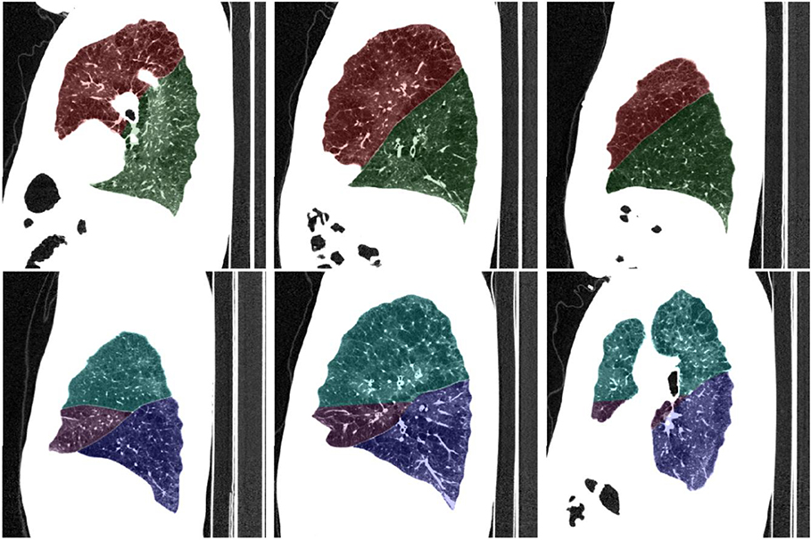
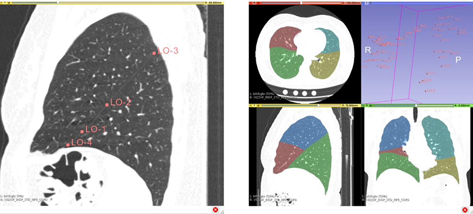
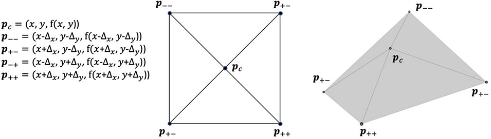
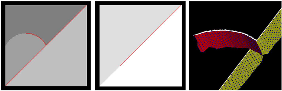
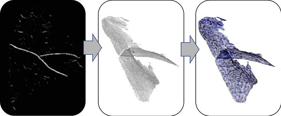
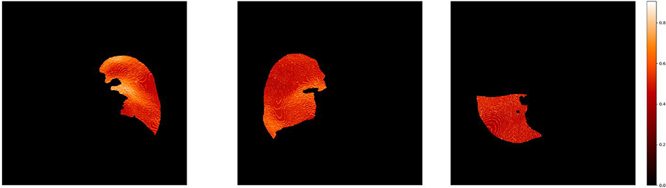
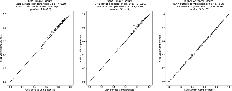
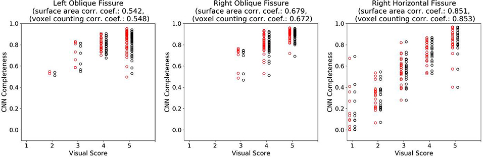
We present an open-source framework for pulmonary fissure completeness assessment. Fissure incompleteness has been shown to associate with emphysema treatment outcomes, motivating the development of tools that facilitate completeness estimation. Generally, the task of fissure completeness assessment requires accurate detection of fissures and definition of the boundary surfaces separating the lung lobes. The framework we describe acknowledges a) the modular nature of fissure detection and lung lobe segmentation (lobe boundary detection), and b) that methods to address these challenges are varied and continually developing. It is designed to be readily deployable on existing lung lobe segmentation and fissure detection data sets. The framework consists of multiple components: a flexible quality control module that enables rapid assessment of lung lobe segmentations, an interactive lobe segmentation tool exposed through 3D Slicer for handling challenging cases, a flexible fissure representation using particles-based sampling that can handle fissure feature-strength or binary fissure detection images, and a module that performs fissure completeness estimation using voxel counting and a novel surface area estimation approach. We demonstrate the usage of the proposed framework by deploying on 100 cases exhibiting various levels of fissure completeness. We compare the two completeness level approaches and also compare to visual reads. The code is available to the community via github as part of the Chest Imaging Platform and a 3D Slicer extension module.
Keywords: Fissure completeness; Lung lobe fissures.
Copyright © 2020 Elsevier Ltd. All rights reserved.
Conflict of interest statement
Declaration of Competing Interest The authors do not hold any conflicts of interest relevant to this manuscript.
Figures
Similar articles
-
A method for the automatic quantification of the completeness of pulmonary fissures: evaluation in a database of subjects with severe emphysema.Eur Radiol. 2012 Feb;22(2):302-9. doi: 10.1007/s00330-011-2278-0. Epub 2011 Oct 8. Eur Radiol. 2012. PMID: 21984417 Free PMC article.
-
Pulmonary lobe segmentation based on ridge surface sampling and shape model fitting.Med Phys. 2013 Dec;40(12):121903. doi: 10.1118/1.4828782. Med Phys. 2013. PMID: 24320514 Free PMC article.
-
Pulmonary Fissure Segmentation in CT Images Using Image Filtering and Machine Learning.Tomography. 2024 Oct 9;10(10):1645-1664. doi: 10.3390/tomography10100121. Tomography. 2024. PMID: 39453038 Free PMC article.
-
When can computed tomography-fissure analysis replace Chartis collateral ventilation assessment in the prediction of patients with emphysema who might benefit from endobronchial valve therapy?Interact Cardiovasc Thorac Surg. 2018 Feb 1;26(2):313-318. doi: 10.1093/icvts/ivx272. Interact Cardiovasc Thorac Surg. 2018. PMID: 29049667 Review.
-
Role of Quantitative Computed Tomographic Scan Analysis in Lung Volume Reduction for Emphysema.Respiration. 2019;98(1):86-94. doi: 10.1159/000498949. Epub 2019 May 8. Respiration. 2019. PMID: 31067563 Review.
Cited by
-
Attention U-net for automated pulmonary fissure integrity analysis in lung computed tomography images.Sci Rep. 2023 Aug 29;13(1):14135. doi: 10.1038/s41598-023-41322-y. Sci Rep. 2023. PMID: 37644125 Free PMC article.
-
A Precise Pulmonary Airway Tree Segmentation Method Using Quasi-Spherical Region Constraint and Tracheal Wall Gap Sealing.Sensors (Basel). 2024 Aug 6;24(16):5104. doi: 10.3390/s24165104. Sensors (Basel). 2024. PMID: 39204799 Free PMC article.
-
Quantifying lung fissure integrity using a three-dimensional patch-based convolutional neural network on CT images for emphysema treatment planning.J Med Imaging (Bellingham). 2024 May;11(3):034502. doi: 10.1117/1.JMI.11.3.034502. Epub 2024 May 29. J Med Imaging (Bellingham). 2024. PMID: 38817711 Free PMC article.
References
-
- Aziz A, Ashizawa K, Nagaoki K, Hayashi K, 2004. High resolution CT anatomy of the pulmonary fissures. J. Thorac. Imaging 19, 186–191. - PubMed
-
- Bookstein FL, 1989. Principal warps: Thin-plate splines and the decomposition of deformations. IEEE Trans. Pattern Anal. Mach. Intell. 11, 567–585.
-
- Delaunay B, others, 1934. Sur la sphere vide. Izv. Akad. Nauk SSSR, Otd. Mat. i Estestv. Nauk 7, 1–2.
-
- Doel T, Gavaghan DJ, Grau V, 2015. Review of automatic pulmonary lobe segmentation methods from CT. Comput. Med. Imaging Graph. 40, 13–29. - PubMed
Publication types
MeSH terms
Grants and funding
LinkOut - more resources
Full Text Sources
