. 2020 Mar 2;9(1):520-533.
doi: 10.1080/22221751.2020.1730709. eCollection 2020.

A chimeric yellow fever-Zika virus vaccine candidate fully protects against yellow fever virus infection in mice

Affiliations

A chimeric yellow fever-Zika virus vaccine candidate fully protects against yellow fever virus infection in mice

Dieudonné Buh Kum et al. Emerg Microbes Infect. .

Abstract

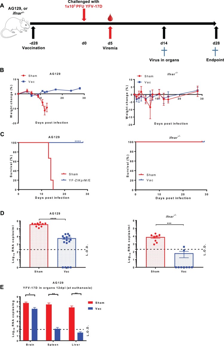
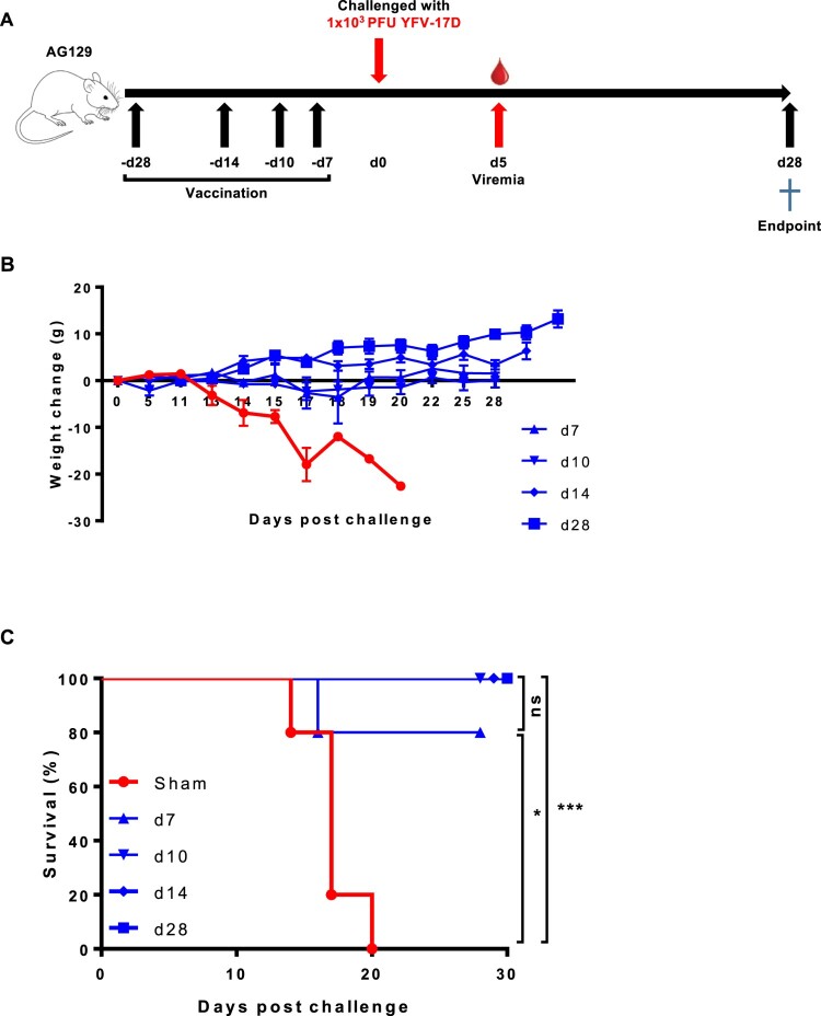
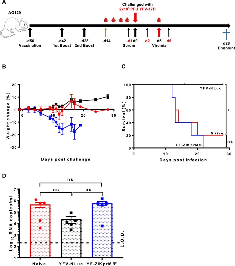
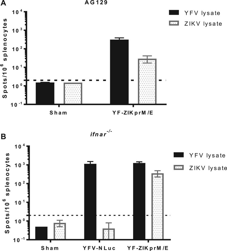
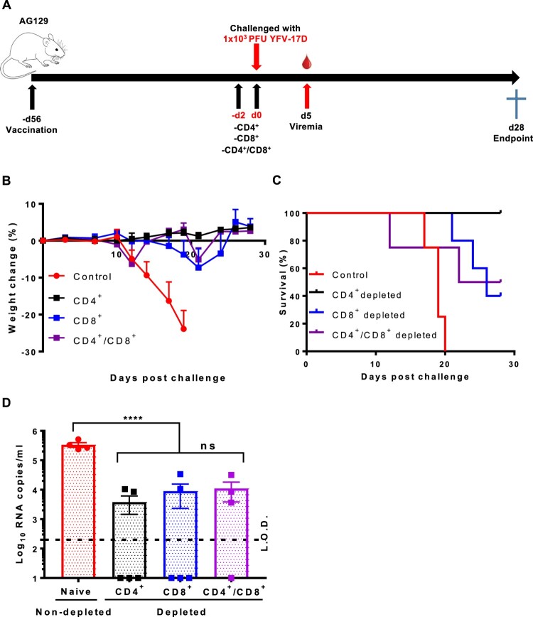
The recent Zika virus (ZIKV) epidemic in the Americas, followed by the yellow fever virus (YFV) outbreaks in Angola and Brazil highlight the urgent need for safe and efficient vaccines against the ZIKV as well as much greater production capacity for the YFV-17D vaccine. Given that the ZIKV and the YFV are largely prevalent in the same geographical areas, vaccines that would provide dual protection against both pathogens may obviously offer a significant benefit. We have recently engineered a chimeric vaccine candidate (YF-ZIKprM/E) by swapping the sequences encoding the YFV-17D surface glycoproteins prM/E by the corresponding sequences of the ZIKV. A single vaccine dose of YF-ZIKprM/E conferred complete protection against a lethal challenge with wild-type ZIKV strains. Surprisingly, this vaccine candidate also efficiently protected against lethal YFV challenge in various mouse models. We demonstrate that CD8+ but not CD4+ T cells, nor ZIKV neutralizing antibodies are required to confer protection against YFV. The chimeric YF-ZIKprM/E vaccine may thus be considered as a dual vaccine candidate efficiently protecting mice against both the ZIKV and the YFV, and this following a single dose immunization. Our finding may be particularly important in the rational design of vaccination strategies against flaviviruses, in particular in areas where YFV and ZIKV co-circulate.

Keywords: CD8+ T cells; YFV-17D; chimeric flavivirus vaccine; live-attenuated vaccines; non-neutralizing antibodies.


Conflict of interest statement

D.B.K, N.M, J.N. and K.D have filed a patent application claiming the discovery and use of chimeric yellow fever-Zika virus vaccines.

Figures

Figure 1.
Figure 1.
Protective efficacy of YF-ZIKprM/E against YFV-17D challenge in AG129 and ifnar−/− mice. (A) Schematic representation of the vaccine-challenge protocol in different mouse models. AG129 and ifnar−/− (6–8 weeks old) were randomly assigned to cages in groups of 5 and either i.p. vaccinated with 1 × 104 PFU of YF-ZIKprM/E (n = 9 or 15) or sham-vaccinated (n = 10). Twenty-eight days after vaccination, mice were challenged with 1 × 103 PFU of YFV-17D. Weight change (B) and survival (C) of sham-vaccinated (red circles) and YF-ZIKprM/E vaccinated (blue squares) mice following i.p. YFV-17D challenge. Challenge virus viremia was quantified by qRT-PCR at day 5 after YFV-17D challenge of AG129 and ifnar−/− mice (D). Red circles represent sham-vaccinated while blue squares represent vaccinated mice. A fraction of asymptomatic vaccinated (n = 5) and symptomatic sham-vaccinated (n = 10) AG129 mice were euthanized and virus titres were quantified by qRT-PCR in the brain, kidney and liver (E). Data are presented as mean values ± SEM from at least 2 independent experiments (n = 5–10). Log-rank (Mantel-Cox) test was used to assess statistical differences in survival rates between sham-vaccinated and vaccinated mice. Mann-Whitney two-tailed test to compare viremia between sham-vaccinated and vaccinated mice. P-values < 0.05 were considered statistically significant. *P < 0.05, **P < 0.01, ***P < 0.001, ****P < 0.0001. Dotted lines denote the limit of detection (L.O.D.) of the assay.
Figure 2.
Figure 2.
Time to protection from YFV-17D-induced weight loss and mortality (i.e. euthanasia) after YF-ZIKprM/E vaccination in AG129 mice. (A) Schematic representation of the time to protection of YF-ZIKprM/E against YFV-17D in AG129 mice. (B) Groups of 6–8 weeks old AG129 mice were either sham-vaccinated (n = 5) or i.p. vaccinated (n = 5/group) with 1 × 104 PFU of YF-ZIKprM/E at different time points (days 28, 14, 10, 7) prior to challenge with 1 × 103 PFU of YFV-17D. Weight and the general condition was monitored for the next 28 days. (C) Survival of sham-vaccinated (red) and YF-ZIKprM/E vaccinated (blue) mice following i.p. challenge with YFV-17D. Data presented as means ± SEM. Log-rank (Mantel-Cox) test was used to assess statistical differences in survival rates between sham-vaccinated and YF-ZIKprM/E vaccinated mice. P-values < 0.05 were considered statistically significant. *P < 0.05, ***P < 0.001, ns = non-significant.
Figure 3.
Figure 3.
Serum from YF-ZIKprM/E vaccinated mice does not neutralize YFV-17D in vitro. Serum samples were collected from sham-vaccinated and YF-ZIKprM/E vaccinated AG129 mice pre- and 14 days post- i.p. challenge with YFV-17D. (A) Total binding antibody (bAb) titres of sham-vaccinated (red circles, n = 10) and YF-ZIKprM/E vaccinated (blue squares, n = 15) sera pre- and post- YFV-17D challenge. (B) Neutralization potential of sham-vaccinated (red circles, n = 10) and YF-ZIKprM/E vaccinated (blue squares, n = 15) sera pre- and post- YFV-17D challenge. Data are presented as mean values ± SEM of at least 2 independent experiments (n = 5–10). Mann-Whitney two-tailed test was performed to assess statistical differences between groups. P-values < 0.05 were considered statistically significant. ***P < 0.001, ****P < 0.0001. Dotted line denotes the limit of detection (L.O.D.) of the assay.
Figure 4.
Figure 4.
Adoptive serum transfer from YF-ZIKprM/E vaccinated mice fails to protect against YFV-17D challenge in AG129 mice. (A) Schematic presentation of adoptive serum transfer in AG129 mice. Mice were either sham-vaccinated (n = 5) or vaccinated with either 1 × 104 PFU of Nanoluciferase expressing YFV-17D (YFV-17D-NLuc) (n = 5) or YF-ZIKprM/E (n = 5) boosted twice with the same doses. Respective sera were collected and pooled and 300 µl of each serum pool injected three times (day −1, 2 and 6) i.p. into 6-weeks old AG129 mice (n = 5 per group) prior to i.p. challenge with 2 × 103 PFU of YFV-17D and a 28 days follow-up. Weight change (B) and survival (C) of AG129 mice that received sera from sham-vaccinated (naive, red circles), YFV-17D-NLuc-vaccinated (black squares) or YF-ZIKprM/E-vaccinated (blue squares). (D) Viremia was quantified by qRT-PCR (5 days post challenge) in mice that received sera from naive (red circles, n = 5), YFV-17D-NLuc (black squares, n = 5) and YF-ZIKprM/E (blue squares, n = 5). Each data point represents a single mouse. Log-rank (Mantel-Cox) test was used to measure statistical differences in survival rates between different groups. Data present mean values with error bars indicating SEM. To compare viremia between groups, two-way ANOVA with Bonferroni correction was used and P-values < 0.05 were considered statistically significant. *P < 0.05. ns = not significant. Dotted line denotes the limit of detection (L.O.D.) of the assay.
Figure 5.
Figure 5.
Ex vivo stimulation of splenocytes from YF-ZIKprM/E and sham-vaccinated mice. AG129 or ifnar−/− mice were either sham-vaccinated or vaccinated with either 1 × 104 PFU of YFV-17D-NLuc or YF-ZIKprM/E. AG129 mice were boosted twice with the same dose (used for vaccination) in intervals of two weeks and later euthanized 10 weeks after the initial vaccination. Ex vivo stimulation of AG129 (A) and ifnar−/− (B) mouse splenocytes with either YFV-17D or ZIKV BeH819015 [29] total cell lysates. Data presenting mean values ± SEM of biologically independent samples (n = 5 per group). Dotted line denotes the limit of detection (L.O.D.) of the assay.
Figure 6.
Figure 6.
CD8+ T cells play a crucial role in the control and clearance of YFV-17D. (A) Schematic representation of T cell depletion in AG129 mice. Mice were either sham-vaccinated (n = 4) or vaccinated with 1 × 104 PFU of YF-ZIKprM/E (n = 4–5/group). Ten weeks after vaccination, vaccinated mice were depleted of either CD4+ (black squares n = 5), CD8+ (blue squares, n = 5) or both (purple squares, n = 4) and challenged i.p. with 1 × 103 PFU of YFV-17D. Weight change (B) and survival (C) was monitored over a period of 28 days following challenge. (D) Viremia was quantified by qRT-PCR 5 days post challenge [CD4+ (black squares, n = 5), CD8+ (blue squares, n = 5) or CD4+/CD8+ depleted (purple squares, n = 4)] presented as mean values ± SEM. To compare viremia between groups, two-way ANOVA with Bonferroni correction was used and P-values < 0.05 were considered statistically significant. ****P < 0.0001. ns = not significant. Dotted line denotes the limit of detection (L.O.D.) of the assay.
Figure 7.
Figure 7.
Cytokine profile of vaccinated and non-vaccinated mice challenged with YFV-17D. Wild-type C57BL/6 or ifnar−/− mice were either sham-vaccinated with MEM 2% FBS or vaccinated with 1 × 104 PFU of YF-ZIKprM/E and challenged 28 days post vaccination. C57BL/6 mice were i.p. administered 2 mg of the MAR1-5A3 1 day prior to challenge. For cytokine measurements, animals were bled by submandibular puncture 5 days post challenge. For YFV-17D challenge, sham-vaccinated (red circles, n = 9) or vaccinated (blue squares, n = 9) ifnar−/−, and sham-vaccinated (red circles, n = 10) or vaccinated (blue squares, n = 10) C57BL/6 were challenged i.p. with 1 × 104 PFU of YFV-17D. Levels of interferon-gamma (A), IL-18 (B), IL-6 (C), TNF-alpha (D), IP-10 (E) and others (Figure S5) were measured in sham-vaccinated (red circles) or vaccinated (blue squares) mice 5 days post challenge. Data presented as means ± SEM. Mann-Whitney two-tailed test was performed to ascertain differences between sham-vaccinated and vaccinated mice. P-values < 0.05 were considered statistically significant. *P < 0.05, **P < 0.01, ***P < 0.001, ****P < 0.0001.

Similar articles

Cited by

References

    1. Fauci AS, Morens DM.. Zika virus in the Americas–Yet another Arbovirus threat. N Engl J Med. 2016;374(7):601–604. doi: 10.1056/NEJMp1600297 - DOI - PubMed
    1. Vasconcelos PF, Monath TP.. Yellow fever remains a potential threat to public health. Vector Borne Zoo Dis. 2016;16(8):566–567. doi: 10.1089/vbz.2016.2031 - DOI - PubMed
    1. Barrett ADT. The reemergence of yellow fever. Science. 2018;361(6405):847–848. doi: 10.1126/science.aau8225 - DOI - PubMed
    1. Shearer FM, Longbottom J, Browne AJ, et al. . Existing and potential infection risk zones of yellow fever worldwide: a modelling analysis. Lancet Glob Health. 2018;6(3):e270–e2e8. doi: 10.1016/S2214-109X(18)30024-X - DOI - PMC - PubMed
    1. Theiler M, Smith HH.. The use of yellow fever virus modified by in vitro cultivation for human immunization. J Exp Med. 1937;65(6):787–800. doi: 10.1084/jem.65.6.787 - DOI - PMC - PubMed

Substances