Review
. 2020 Feb 7:12:329.
doi: 10.3389/fnmol.2019.00329. eCollection 2019.

Zebrafish as a Model System for the Study of Severe CaV2.1 (α1A) Channelopathies

Affiliations
Review

Zebrafish as a Model System for the Study of Severe CaV2.1 (α1A) Channelopathies

Sidharth Tyagi et al. Front Mol Neurosci. .

Abstract

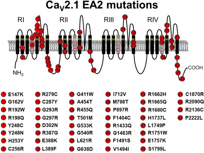
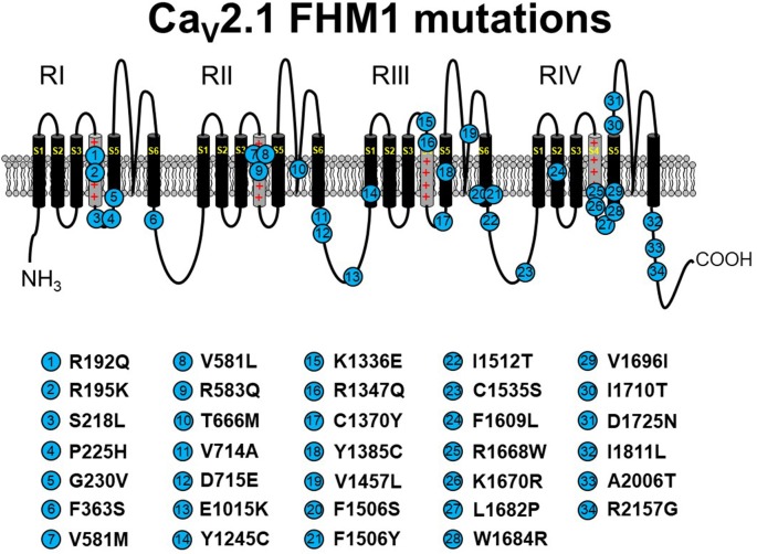
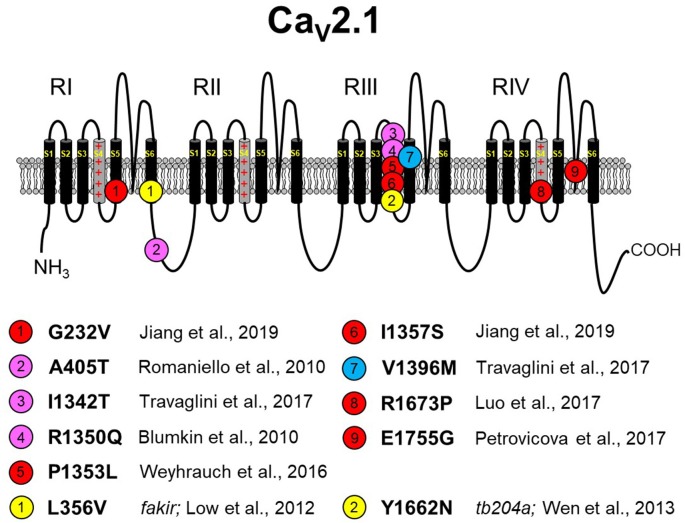
The P/Q-type CaV2.1 channel regulates neurotransmitter release at neuromuscular junctions (NMJ) and many central synapses. CACNA1A encodes the pore-containing α1A subunit of CaV2.1 channels. In humans, de novo CACNA1A mutations result in a wide spectrum of neurological, neuromuscular, and movement disorders, such as familial hemiplegic migraine type 1 (FHM1), episodic ataxia type 2 (EA2), as well as a more recently discovered class of more severe disorders, which are characterized by ataxia, hypotonia, cerebellar atrophy, and cognitive/developmental delay. Heterologous expression of CaV2.1 channels has allowed for an understanding of the consequences of CACNA1A missense mutations on channel function. In contrast, a mechanistic understanding of how specific CACNA1A mutations lead in vivo to the resultant phenotypes is lacking. In this review, we present the zebrafish as a model to both study in vivo mechanisms of CACNA1A mutations that result in synaptic and behavioral defects and to screen for effective drug therapies to combat these and other CaV2.1 channelopathies.

Keywords: CaV2.1; P/Q-type; channelopathy; episodic ataxia type 2; familial hemiplegic migraine type 1; vertebrate models; zebrafish; α1A.


Figures

Figure 1
Figure 1
Schematic representation of human CaV2.1 mutations causing episodic ataxia type 2 (EA2). Please note that residue numbering varies between studies due to the existence of multiple CACNA1A splice variants; residue numbers indicated reflect those stated in the original report. Citations to the indicated mutations are listed as follows: E147K—Imbrici et al., ; G162V—Maksemous et al. (2016); R192W—Soden et al. (2014); R198Q—Indelicato et al. (2019); Y248C—Zafeiriou et al. (2009); Y248N—Choi et al. (2017); H253Y—van den Maagdenberg et al. (2002); C256R—Mantuano et al. (2004); R279C—Maksemous et al. (2016); C287Y—Jen et al. (2004); G293R—Yue et al. (1997); G297R—Tantsis et al. (2016); D302N—Maksemous et al. (2016); R387G—Maksemous et al. (2016); E388K—Nikaido et al. (2011); L389F—Mantuano et al. (2010); G411W—Maksemous et al. (2016); A454T—Cricchi et al. (2007); R455Q—Isaacs et al. (2017); T501M—Mantuano et al. (2010); G533K—Scoggan et al. (2006); G540R—Rajakulendran et al. (2010a); L621R—Rajakulendran et al. (2010a); G638D—Cuenca-León et al. (2009); I712V—Guerin et al. (2008); M798T—Mantuano et al. (2010); P897R—Mantuano et al. (2010); F1404C—Jen et al. (2001); R1433Q—Pietrobon (2010); G1483R—Mantuano et al. (2004); F1491S—Guida et al. (2001); V1494I—Mantuano et al. (2004); R1662H—Friend et al. (1999); R1665Q—Tonelli et al. (2006); R1680C—Mantuano et al. (2010); H1737L—Spacey et al. (2004); L1749P—Maksemous et al. (2016); R1751W—Bertholon et al. (2009); E1757K—Denier et al. (2001); S1799L—Ohba et al. (2013); C1870R—Mantuano et al. (2010); R2090Q—Melzer et al. (2010); R2136C—Mantuano et al. (2004); P2222L—Sintas et al. (2017). The CaV2.1 schematic was modified from Tyagi et al. (2019) with permission of the authors.
Figure 2
Figure 2
Schematic representation of human CaV2.1 mutations causing familial hemiplegic migraine type 1 (FHM1). Please note that residue numbering varies between studies due to the existence of multiple CACNA1A splice variants; residue numbers indicated reflect those stated in the original report. Citations to the indicated mutations are listed as follows: R192Q—Ophoff et al. (1996); R195K—Ducros et al. (2001); S218L—Kors et al. (2001); P225H—Stuart et al. (2012); G230V—Yang et al. (2014); F363S—Riant et al. (2010); V581M—Cuenca-León et al. (2008); V581L—Freilinger et al. (2011); R583Q—Battistini et al. (1999); T666M—Ophoff et al. (1996); V714A—Ophoff et al. (1996); D715E—Ducros et al. (2001); E1015K—Grieco et al. (2018); Y1245C—Cuenca-León et al. (2008); K1336E—Ducros et al. (2001); R1347Q—Alonso et al. (2004); C1370Y—Thomsen et al. (2007); Y1385C—Vahedi et al. (2000); V1457L—Carrera et al. (1999); F1506S—Riant et al. (2010); F1506Y—Pelzer et al. (2018); I1512T—Grieco et al. (2018); C1535S—Dichgans et al. (2005); F1609L—Pelzer et al. (2018); R1668W—Ducros et al. (2001); K1670R—Riant et al. (2010); L1682P—Weiss et al. (2007); W1684R—Ducros et al. (2001); V1696I—Ducros et al. (2001); I1710T—Kors et al. (2004); D1725N—Riant et al. (2010); I1811L—Ophoff et al. (1996); A2006T—Wilson (2014); R2157G—Grieco et al. (2018). The CaV2.1 schematic was modified from Tyagi et al. (2019) with permission of the authors.
Figure 3
Figure 3
Missense CaV2.1 mutations leading to neurodevelopmental disorders. The zebrafish fakir and tb204a mutants are also depicted as yellow circles. Red circles indicate a loss-of-function human mutation. Blue circles indicate a gain-of-function human mutation. Magenta circles indicate a yet-to-be functionally characterized human mutation. Specific references are indicated below. As in Figures 1, 2, please note that residue numbering varies between studies due to: (1) the existence of multiple known CACNA1A splice variants; and (2) species differences between humans and zebrafish. The CaV2.1 schematic was modified from Tyagi et al. (2019) with permission of the authors. These mutations are discussed in sections “The Expanding Spectrum OF CaV2.1-α1A Channelopathies” and “Zebrafish as a Model SYSTEM for the Study of Severe CaV2.1 Channelopathies.”

References

    1. Adams P. J., Garcia E., David L. S., Mulatz K. J., Spacey S. D., Snutch T. P. (2009). CaV2.1 P/Q-type calcium channel alternative splicing affects the functional impact of familial hemiplegic migraine mutations: implications for calcium channelopathies. Channels 3, 110–121. 10.4161/chan.3.2.7932 - DOI - PubMed
    1. Aggarwal S. K., MacKinnon R. (1996). Contribution of the S4 segment to gating charge in the Shaker K+ channel. Neuron 16, 1169–1177. 10.1016/s0896-6273(00)80143-9 - DOI - PubMed
    1. Alonso I., Barros J., Tuna A., Seixas A., Coutinho P., Sequeiros J., et al. . (2004). A novel R1347Q mutation in the predicted voltage sensor segment of the P/Q-type calcium-channel α-subunit in a family with progressive cerebellar ataxia and hemiplegic migraine. Clin. Genet. 65, 70–72. 10.1111/j..2004.00187.x - DOI - PubMed
    1. Althobaiti Y. S., Almalki A., Alsaab H., Alsanie W., Gaber A., Alhadidi Q., et al. . (2019). Pregabalin: potential for addiction and a possible glutamatergic mechanism. Sci. Rep. 9:15136. 10.1038/s41598-019-51556-4 - DOI - PMC - PubMed
    1. Bahamonde M. I., Serra S. A., Drechsel O., Rahman R., Marcé-Grau A., Prieto M., et al. . (2015). A single amino acid deletion (ΔF1502) in the S6 segment of CaV2.1 domain III associated with congenital ataxia increases channel activity and promotes Ca2+ influx. PLoS One 10:e0146035. 10.1371/journal.pone.0146035 - DOI - PMC - PubMed

LinkOut - more resources